
Medical History: Anatomy Models, from Antique to Modern-Day
Nov 6th 2025
Derived from the Greek words ἀνατομή anatomē (“to cut open”), anatomy is medicine’s oldest scientific discipline. Central to its teaching are anatomical models – three-dimensional representations of the human body or its parts, created to help visualize structures that are difficult to study in living subjects. For centuries, anatomical models have shaped how we study the body, evolving from handcrafted antique anatomical models to today’s technology-driven, lifelike simulation tools.
For educators, students, and medical professionals alike, understanding the rich history of anatomical models illuminates how advances in science, craftsmanship, and technology work in harmony to transform critical medical procedures.
The Role of 3D Anatomical Models in Medical Education
Before diving into the historical progression of anatomy models, it’s essential to recognize why anatomical models have been integral to medical education. Unlike early 2D illustrations and written descriptions, these three-dimensional tools offer tactile and spatial understanding, helping learners grasp the complexity of bones, organs, and systems. They serve as safer, sustainable alternatives to cadavers and give students an opportunity to practice concepts.
What Materials Were Anatomical Models Made of Historically?
Artisanship and material innovations have shaped how models are used. Original models were handmade with wood, bronze, and other locally available resources. Antique models were handmade with wax, papier-mâché, resin, and more. Today’s models are made of plastics and metals using 3D printing. Each new material and manufacturing improvement has enhanced anatomy models’ durability, accuracy, and accessibility for students, educators, and medical professionals. This fascinating evolution underscores how models not only mirror scientific progress but also reflect changing cultural and technological priorities in medicine.
With that foundation in mind, let’s explore the timeline of how anatomical models have evolved across centuries.
The Evolution of Anatomy Models: From the 18th Century to the Digital Age
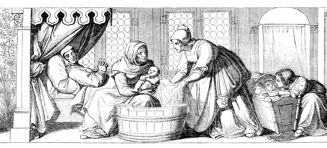
18th Century: Wax & Obstetrics
During the 1700s, dissection opportunities were rare, making anatomy models critical to medical education. Obstetrics models in the 18th century were among the first widely adopted teaching tools, often crafted from wax, which could be easily dyed and molded with a high level of detail. These models helped midwives and physicians study childbirth without waiting for real births or studying cadavers. Because these 18th-century models were handmade, each piece was unique and often considered a work of art as much as an educational tool. Today, collectors and historians prize these antique anatomical models for their historical and artistic value.
Key Features
- Focused on childbirth and midwifery training
- Individually crafted by hand and therefore unique
- Both scientific tools and artistic works
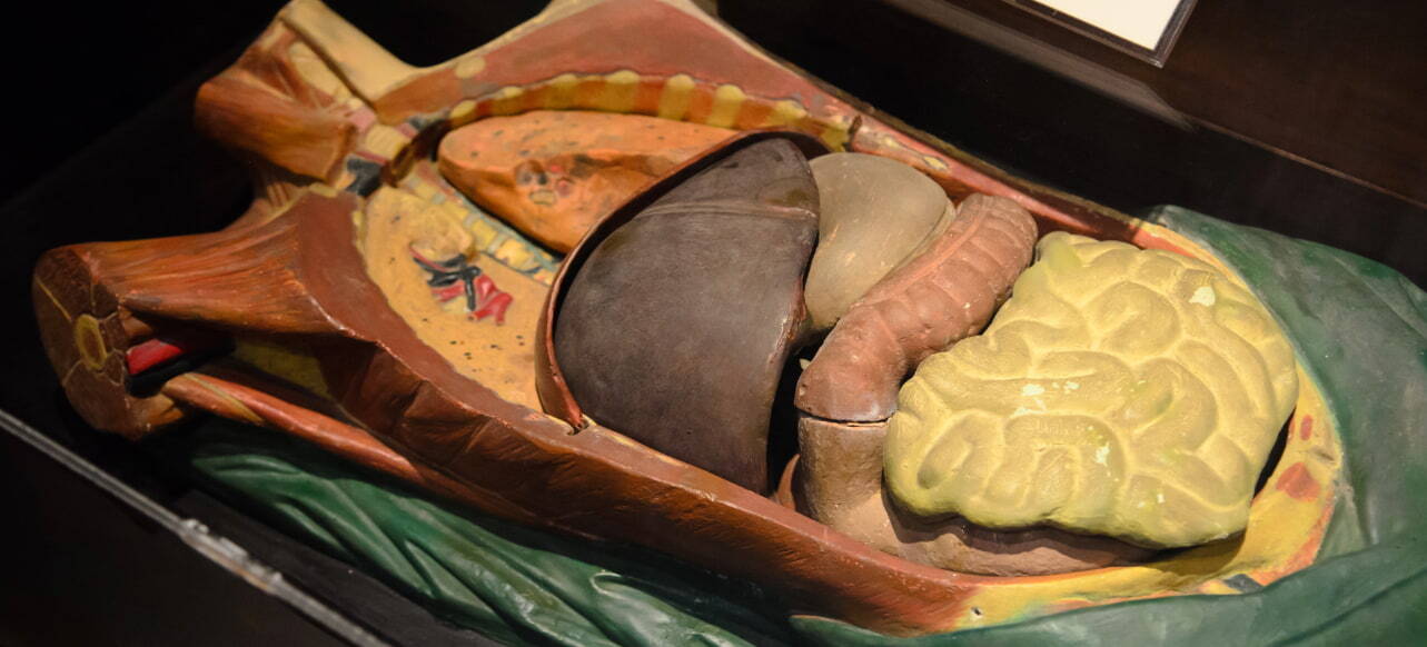
19th Century: Expansion & Craftsmanship
European manufacturers, particularly in Italy and Germany, refined the process of wax modeling in the 1800s. Additionally, papier-mâché emerged as a more affordable material for making vintage anatomical models. By this period, students and doctors could learn from large, highly detailed models of the human skeleton, muscles, and organs. These models were not only durable but portable, too. They were often engineered with removable parts to aid instruction through authentic simulations. By making anatomy accessible to a wider audience, these 19th-century models paved the way for the mass production of anatomical models at the turn of the 20th century.
Key Features
- More affordable materials
- Quicker manufacturing and wider distribution
- Greater detail in musculoskeletal representations and internal organ models
- Expanded access to vital medical knowledge beyond elite medical schools
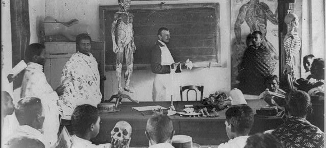
Early 20th Century: Industrial Manufacturing
As industrialization spread, anatomical model manufacturers began producing standardized models on a large scale. Resin, plaster, and early plastics replaced fragile wax and papier-mâché materials. This manufacturing shift allowed for greater durability and mass distribution, making models available not only to universities but also to secondary schools and public health initiatives, like Germany’s International Hygiene Exhibition of 1911. These models also became increasingly precise thanks to advances in dissection practices and medical research.
Key Factors
- More standardized
- More durable due to material innovations
- Growing global accessibility
- Anatomical precision improved through research advances
Mid-20th Century: The Plastics Revolution
By the 1950s and 1960s, plastics became the primary material for anatomical models. Most mid-20th-century models were composed of PVC, acrylic, or other polymers that could withstand frequent handling. These lightweight, low-cost materials meant that models could now be produced in bulk, shipped globally, and incorporated into classrooms of all sizes and grade levels. This further democratized medical education, allowing aspiring doctors, nurses, and allied health professionals to engage with state-of-the-art learning tools.
Key Factors
- Cost-effective due to mass manufacturing
- Durable and portable for frequent handling
- Democratized access to anatomical education and necessary learning aids
21st Century: Digital & Simulation Inventions
Today’s anatomical models combine craftsmanship with cutting-edge technological innovation. Hyper-realistic models mimic the look and feel of human tissue and organs. Advanced healthcare simulation models incorporate virtual reality systems that “breathe,” respond to touch, and display changes in vital signs. These innovations not only prepare students for real-life scenarios but also expand access to training.
Key Factors
- Hyper-realistic models supporting training for specific procedures and emergencies
- Integrated digital features
- Expanded training (e.g., in areas where cadaver access is limited)
Why Anatomical Model Evolution Matters
From antique anatomical models carved in wax to today’s machine-learning-powered simulators, the progression of anatomical models mirrors the progress of medicine itself. Each innovation has solved critical challenges in education and public health: overcoming scarcity, improving access, and guiding ground-breaking medical advancements worldwide. For educators and students, these tools remain essential for bridging theory and practice.
As technology continues to evolve, so will the ways we learn about the human body. Whether you’re an educator equipping your classroom or a student seeking study tools, explore Anatomy Warehouse’s modern models that reflect centuries of progress. Feel free to contact us for guidance or request a custom quote to achieve your learning objectives.
Share:


