Description
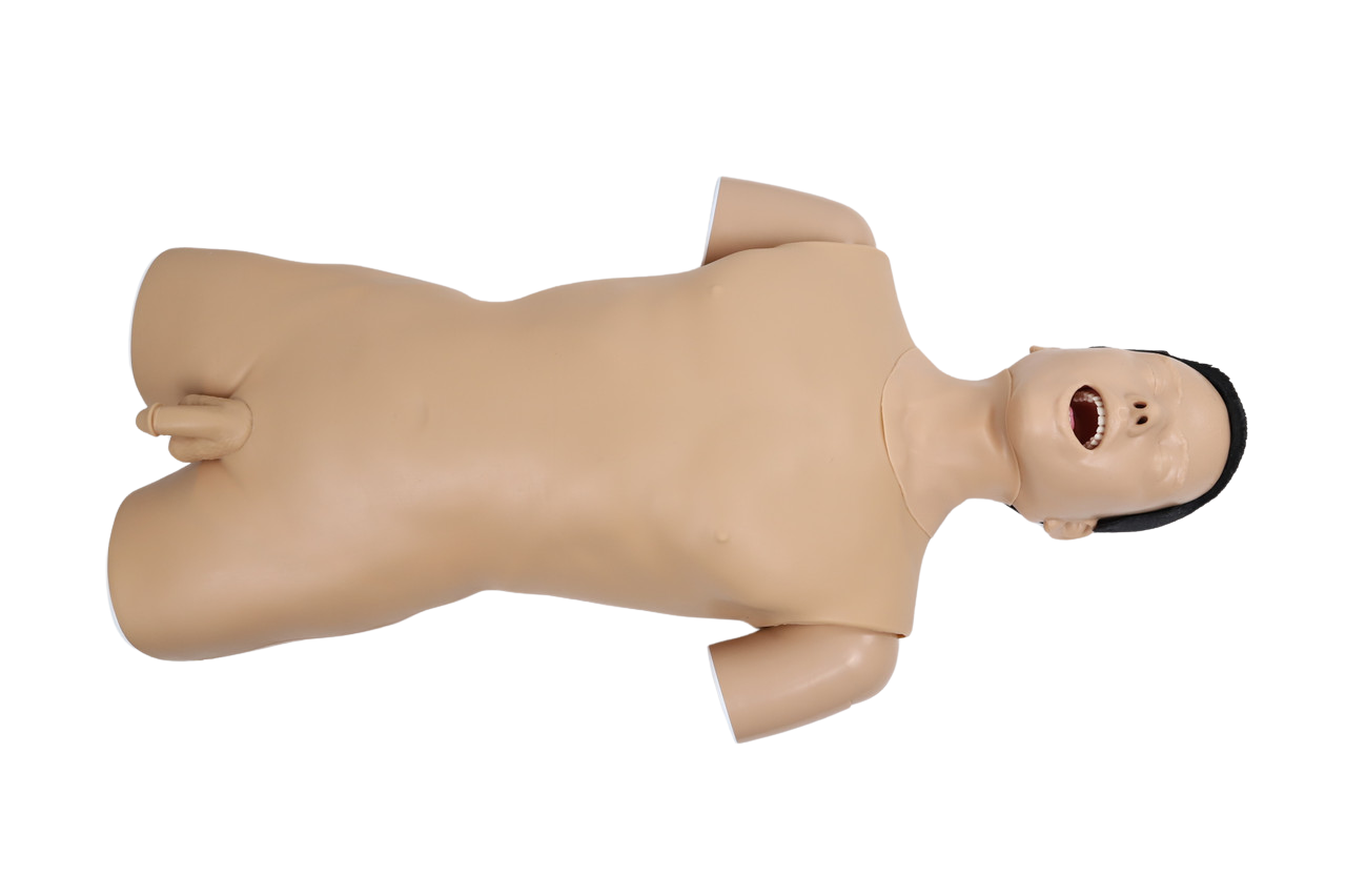
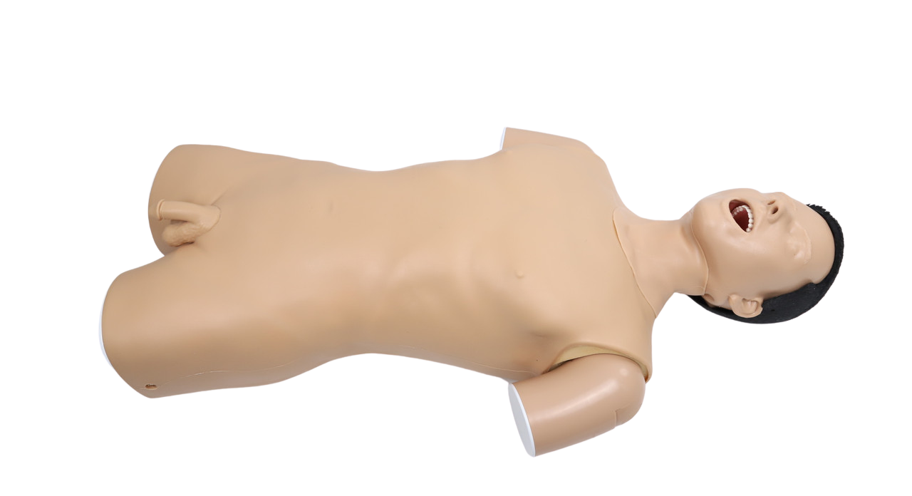
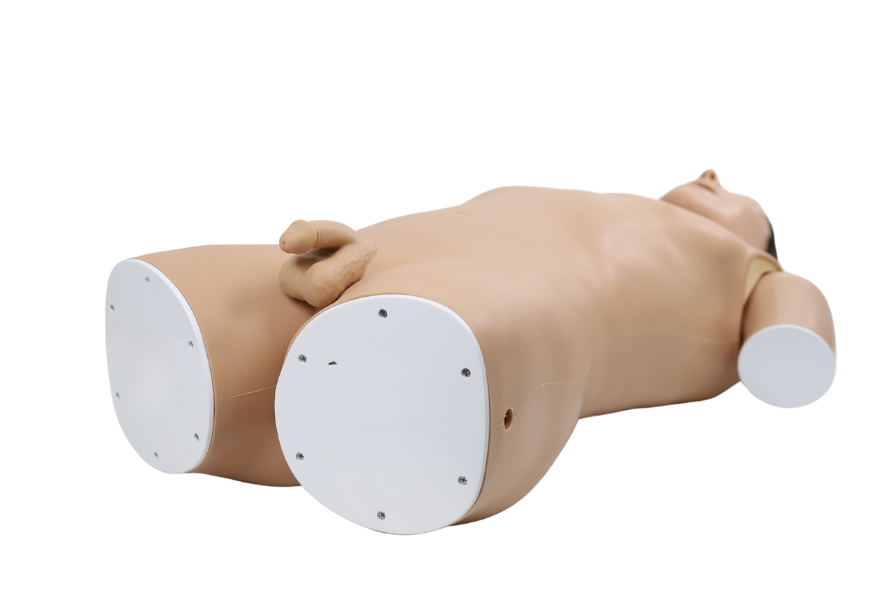
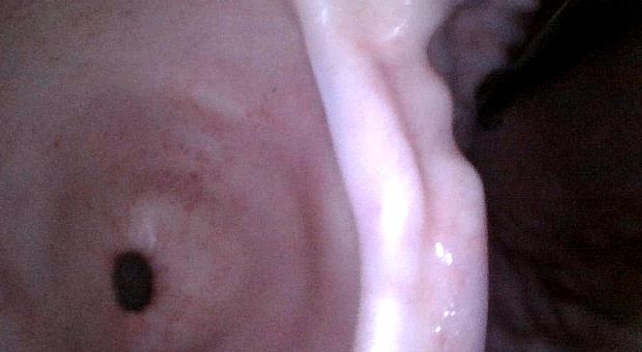
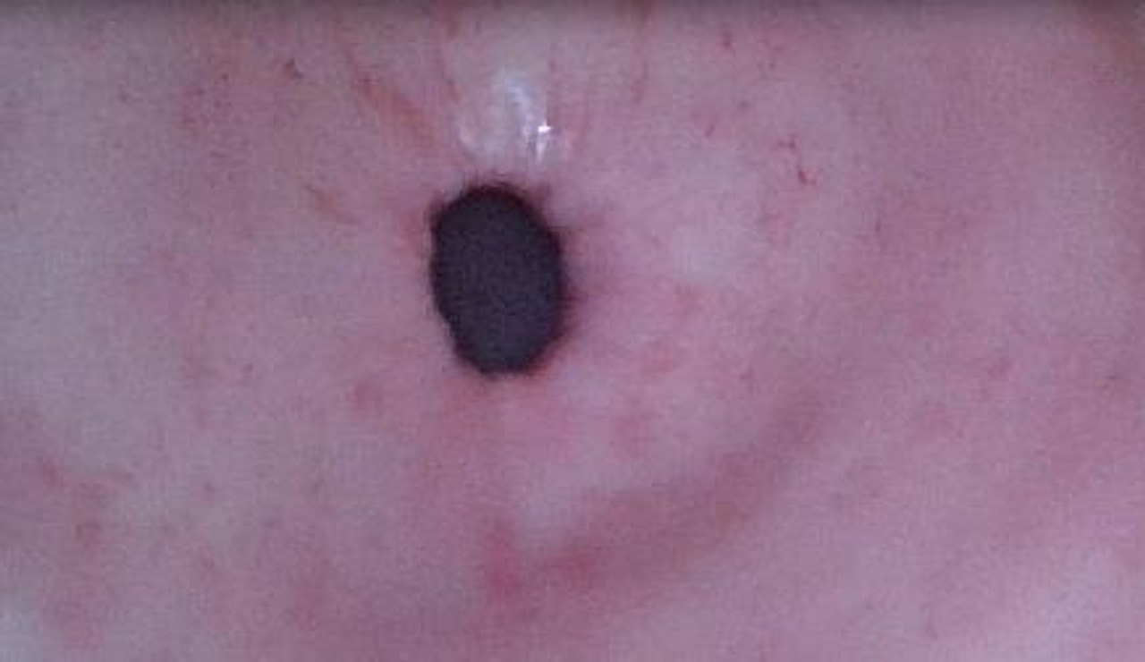
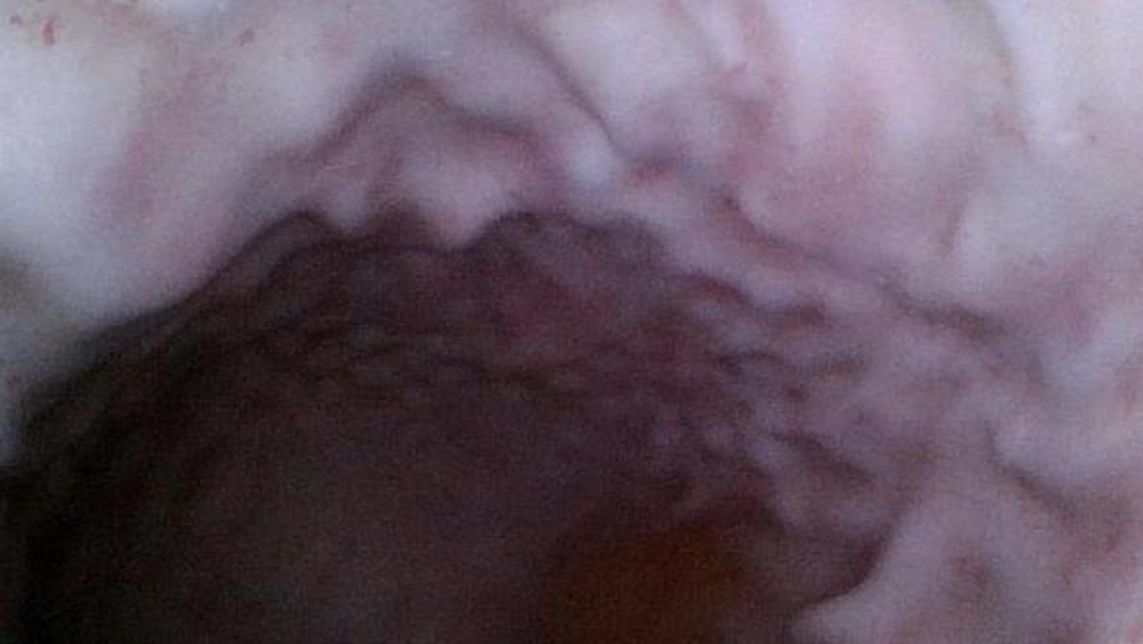
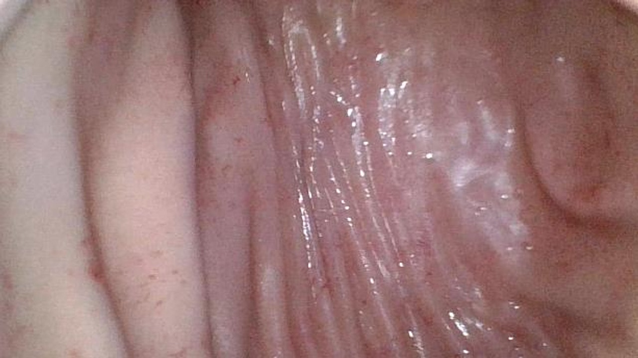
Upper GI Endoscopy Training Model
Skills Gained
- Understanding of anatomical structures
- Fiberoptic endoscopy
- ERCP
- Endoscopy
Features
Anatomy
- Nasal cavity
- Oral cavity
- Pharynx
- Tonsil
- Palatoglossal arch
- Velopharyngeal arch
- Pyriform recess
- Epiglottis
- Tracheal opening
- Esophageal opening
- Esophagus
- Stomach
- Duodenum
Realism
- Realistic male appearance including head, neck, and torso
- CT scan of a real person’s upper digestive tract
Key Features
- Realistic and complete internal structures including:
- Cardia
- Dentate line
- Pylorus
- Major duodenal papilla (papilla of Vater)
- Accessory duodenal papilla
- Gastric angle
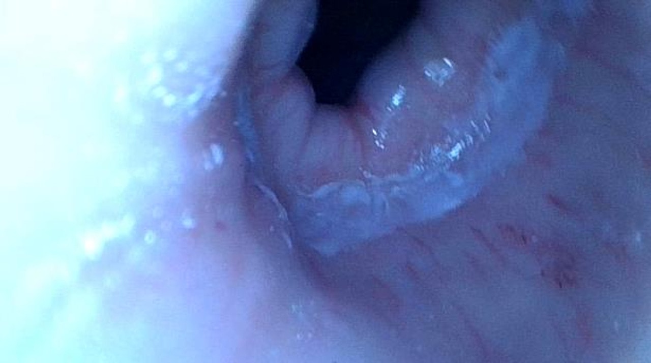
- Use endoscope to observe mucosa, showing lifelike mucosa color, shape, and feel
- The stomach can be pumped and inflated

