Description
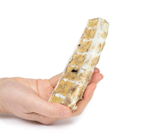
Developed from real patient case study specimens, the 3D printed anatomy model pathology series introduces an unmatched level of realism in human anatomy models. Each 3D printed anatomy model is a high-fidelity replica of a human cadaveric specimen, focusing on the key morbidity presentations that led to the deceasement of the patient. With advances in 3D printing materials and techniques, these stories can come to life in an ethical, consistently reproduceable, and easy to handle format. Ideal for the most advanced anatomical and pathological study, and backed by authentic case study details, students, instructors, and experts alike will discover a new level of anatomical study with the 3D printed anatomy model pathology series.
Clinical History
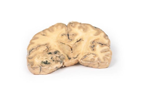
A 89-year old male presents with an episode of large hemoptysis. He has a history of diabetes and immunosuppression secondary to steroid treatment for rheumatoid arthritis. Further history reveals a long history of cough, hemoptysis, fevers and weight loss. On examination, he is noted to be cachexic, hypoxic and have crepitations throughout the left lung. Chest x-ray shows multiple cavitation lesions in the left lung. Subsequently, he has another massive hemoptysis and dies.
Pathology
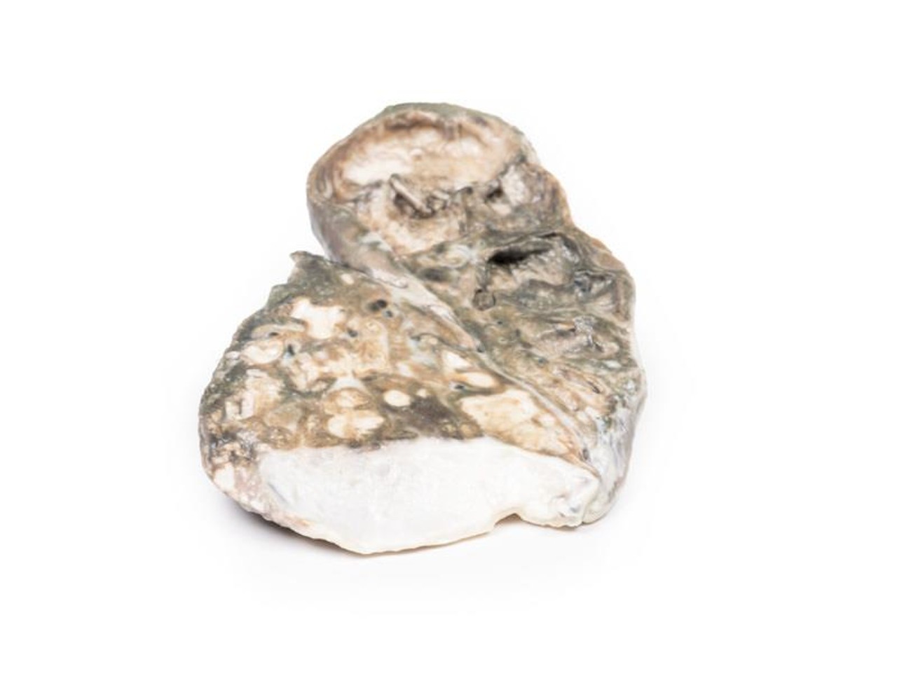
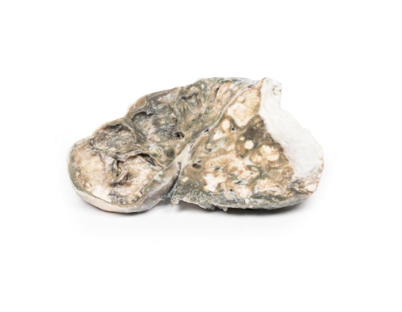
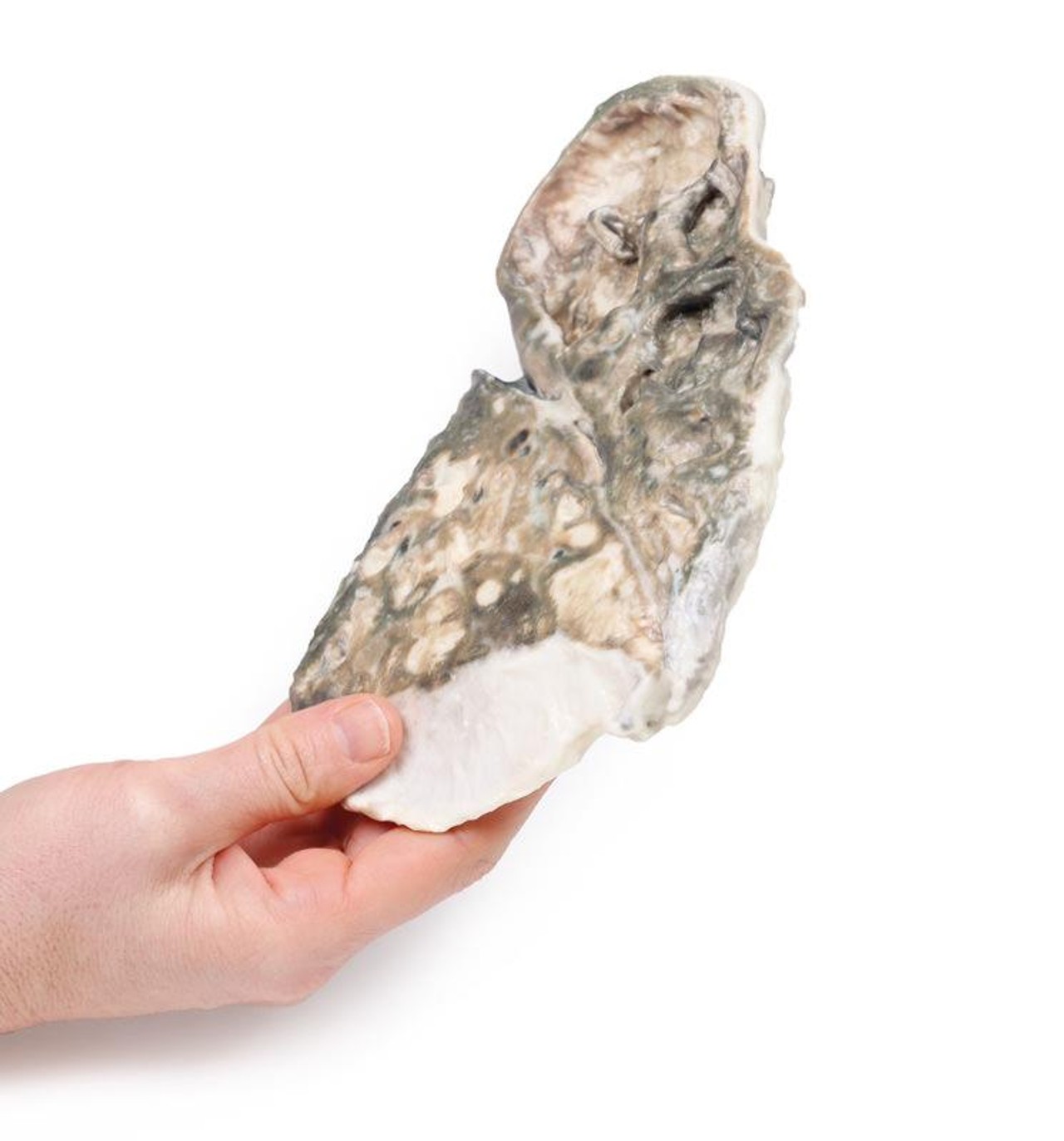
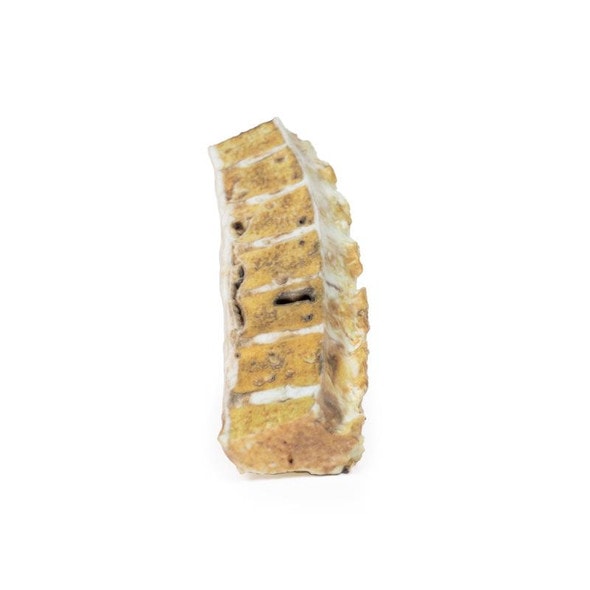
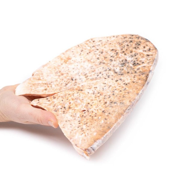
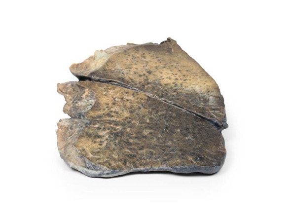
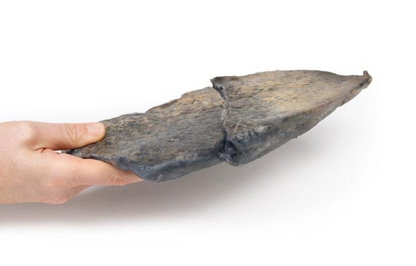
The left lung is cut longitudinally to display the cut surface. The upper lobe is almost entirely replaced by several large irregular cavities lined by necrotic debris and fibrous tissue. Blood vessels are seen in the upper cavity with evidence of hemorrhage. The lower lobe contains several smaller caseous areas, some of which are breaking down. The intervening lung parenchyma is scarred. The pleura is thickened. This is fibrocaseous tuberculosis with cavitation.
Further Information
Tuberculosis (TB) is a chronic pulmonary and systemic infectious disease caused by Mycobacteria tuberculosis. Transmission most commonly occurs via inhalation of aerosolized droplets of M. tuberculosis. Risk factors for contracting TB include being an inhabitant of a developing country where the disease may be endemic, immunosuppression (e.g. HIV, steroid use, anti-TNF use and diabetes), chronic lung disease (e.g. silicosis), alcoholism and malnutrition.
After initial pulmonary infection of M. tuberculosis clinical manifestation varies. In 90% of individuals with an intact immune system they enter an asymptomatic latent infection phase. This latent TB may reactivate at any time in the patient's life. In the other 10% of patients, especially in the immunocompromised, they develop primary disease which is immediate active TB infection. Manifestations of primary TB include pulmonary infection symptoms (e.g. consolidation, effusion and hilar adenopathy) and extra pulmonary symptoms including lymphadenopathy, meningitis and disseminated miliary TB.
Secondary tuberculosis occurs when there is reactivation of previous latent TB infection. Around 10% of latent TB will reactivate usually during periods of weakened host immunity. Typical symptoms of reactivation are cough, hemoptysis, low grade fever, night sweats and weight loss.
The immune response against TB is mediated via TH1-cells stimulate alveolar macrophages to attack the mycobacteria. These macrophages surround the infection forming a granuloma with central caseous necrosis.
Secondary pulmonary TB may heal with fibrosis or progress as in this case. Progressive pulmonary TB sees erosion and expansion of the infectious lesion into adjacent lung parenchyma. This leads to evacuation of the caseous center leading to fibrous cavitation. Erosion of blood vessels can occur causing hemoptysis. Post treatment of TB the tissue heals by fibrosis but does not recover the pulmonary architecture.
TB diagnosis is usually made with a clinical history and chest x-ray and multiple sputum cultures. Mantoux skin tuberculin test and serum interferon gamma release assay may also be used to help screen for infection. Biopsies may be taken of suspected infection site for culture to assist diagnosis. Treatment involves prolonged courses of multiple antibiotics, which depend on the antibiotic resistance of the infecting mycobacterium.
Advantages of 3D Printed Anatomical Models
- 3D printed anatomical models are the most anatomically accurate examples of human anatomy because they are based on real human specimens.
- Avoid the ethical complications and complex handling, storage, and documentation requirements with 3D printed models when compared to human cadaveric specimens.
- 3D printed anatomy models are far less expensive than real human cadaveric specimens.
- Reproducibility and consistency allow for standardization of education and faster availability of models when you need them.
- Customization options are available for specific applications or educational needs. Enlargement, highlighting of specific anatomical structures, cutaway views, and more are just some of the customizations available.
Disadvantages of Human Cadavers
- Access to cadavers can be problematic and ethical complications are hard to avoid. Many countries cannot access cadavers for cultural and religious reasons.
- Human cadavers are costly to procure and require expensive storage facilities and dedicated staff to maintain them. Maintenance of the facility alone is costly.
- The cost to develop a cadaver lab or plastination technique is extremely high. Those funds could purchase hundreds of easy to handle, realistic 3D printed anatomical replicas.
- Wet specimens cannot be used in uncertified labs. Certification is expensive and time-consuming.
- Exposure to preservation fluids and chemicals is known to cause long-term health problems for lab workers and students. 3D printed anatomical replicas are safe to handle without any special equipment.
- Lack of reuse and reproducibility. If a dissection mistake is made, a new specimen has to be used and students have to start all over again.
Disadvantages of Plastinated Specimens
- Like real human cadaveric specimens, plastinated models are extremely expensive.
- Plastinated specimens still require real human samples and pose the same ethical issues as real human cadavers.
- The plastination process is extensive and takes months or longer to complete. 3D printed human anatomical models are available in a fraction of the time.
- Plastinated models, like human cadavers, are one of a kind and can only showcase one presentation of human anatomy.
Advanced 3D Printing Techniques for Superior Results
- Vibrant color offering with 10 million colors
- UV-curable inkjet printing
- High quality 3D printing that can create products that are delicate, extremely precise, and incredibly realistic
- To improve durability of fragile, thin, and delicate arteries, veins or vessels, a clear support material is printed in key areas. This makes the models robust so they can be handled by students easily.

