Description
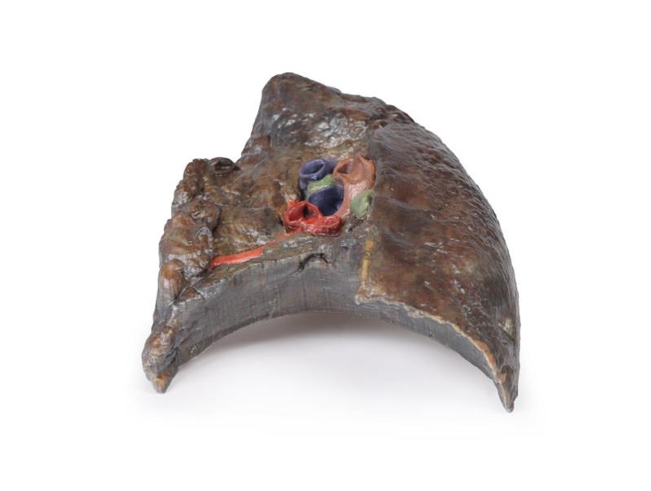
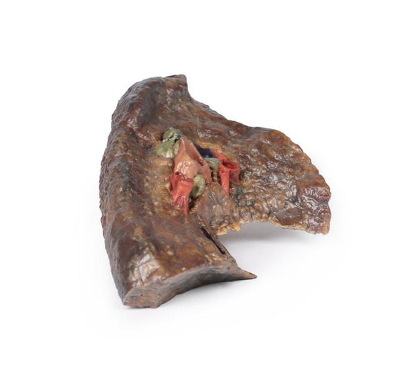
The hilum of a lung is the point at which visceral and parietal pleura meet and functions with the pulmonary ligament as the lungs only connection with the rest of the body. This connection includes the Pulmonary Artery, Superior and Inferior Pulmonary Veins, Main Bronchi, Nerves and Lymphatics.
As the definition of an artery involves carrying blood AWAY from the heart, this will be deoxygenated blood in the pulmonary system, in contrast with the systemic circulation. Similarly, veins carry blood TOWARDS the heart, meaning it will be oxygenated in the pulmonary system.
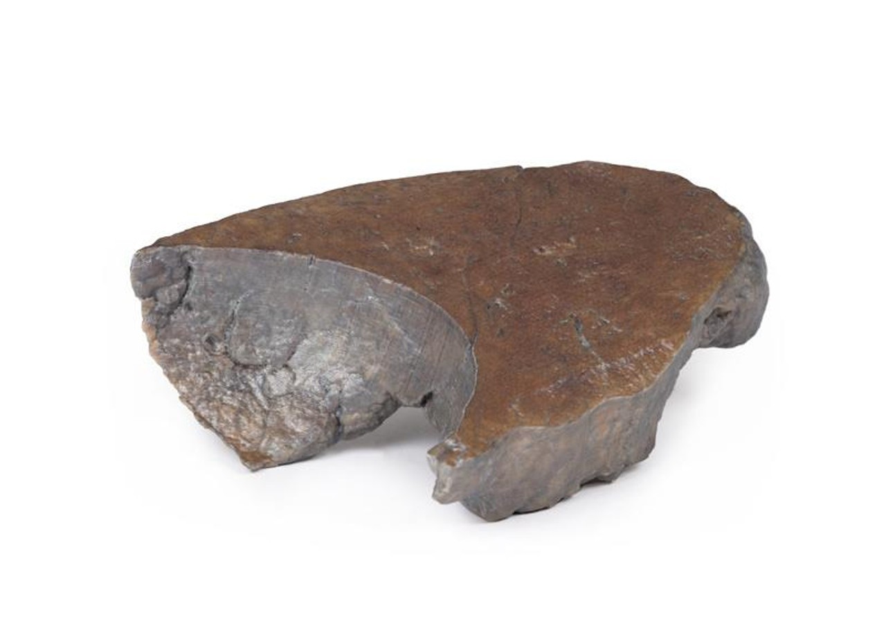
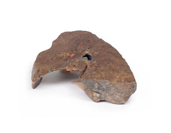
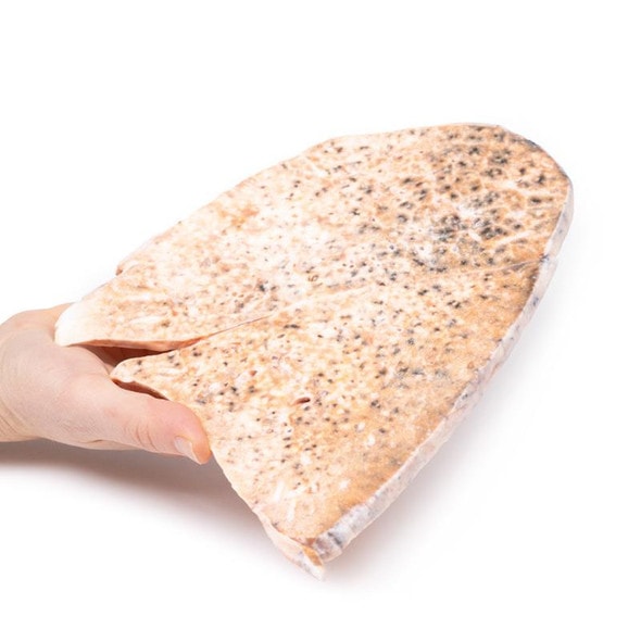
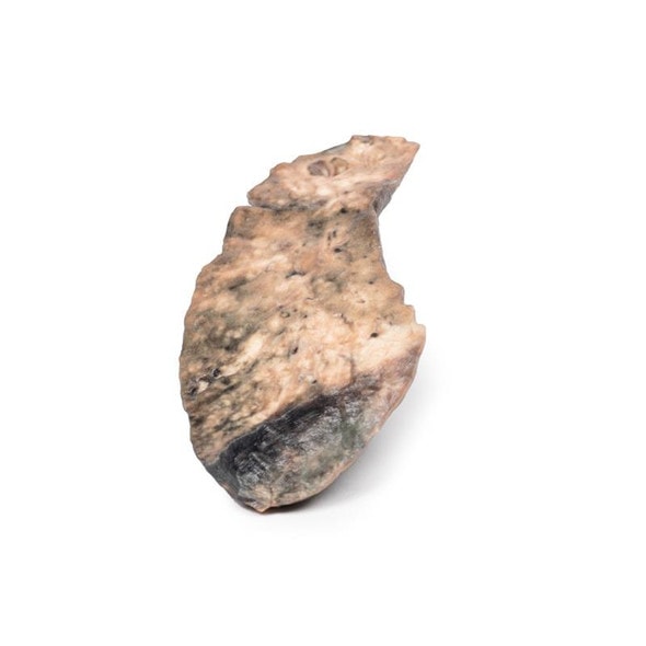
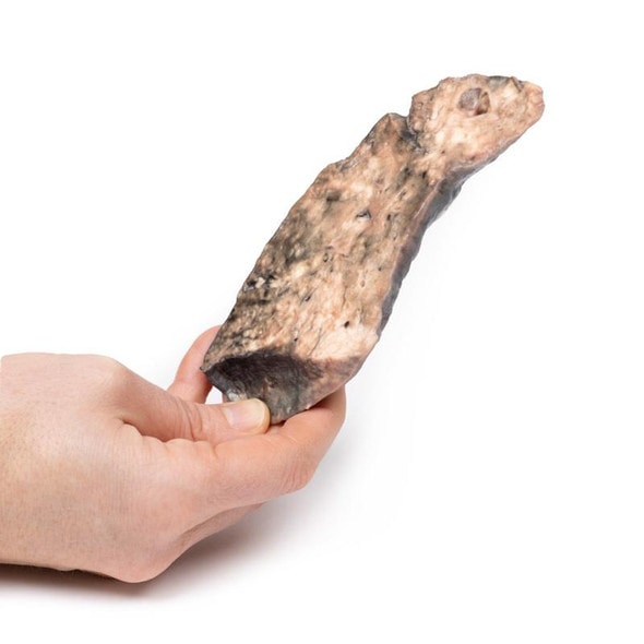
With the specimen cut in a sagittal plane in line with the cardiac impression, nerves and lymphatics are difficult to identify however the groove from the oesophagus as it descends posteriorly to pierce the diaphragm can be seen alongside the cardiac impression (of the right atrium) is notable anterior to the hilum of the right lung; the right main bronchi and its subsequent divisions into lobar bronchi, found in this specimen more posterior in the hilum; he pulmonary artery and its divisions, located most superior within the hilum; the superior and inferior pulmonary veins and their divisions which are most inferior and anterior in the specimen. the oblique and horizontal fissures along the lateral surface of the specimen and the Hilar lymph nodes around the hilum on the medial surface of the lung.
The diaphragmatic surface is found inferiorly and the costal visceral surface is on the posterior of the specimen.
Advantages of 3D Printed Anatomical Models
- 3D printed anatomical models are the most anatomically accurate examples of human anatomy because they are based on real human specimens.
- Avoid the ethical complications and complex handling, storage, and documentation requirements with 3D printed models when compared to human cadaveric specimens.
- 3D printed anatomy models are far less expensive than real human cadaveric specimens.
- Reproducibility and consistency allow for standardization of education and faster availability of models when you need them.
- Customization options are available for specific applications or educational needs. Enlargement, highlighting of specific anatomical structures, cutaway views, and more are just some of the customizations available.
Disadvantages of Human Cadavers
- Access to cadavers can be problematic and ethical complications are hard to avoid. Many countries cannot access cadavers for cultural and religious reasons.
- Human cadavers are costly to procure and require expensive storage facilities and dedicated staff to maintain them. Maintenance of the facility alone is costly.
- The cost to develop a cadaver lab or plastination technique is extremely high. Those funds could purchase hundreds of easy to handle, realistic 3D printed anatomical replicas.
- Wet specimens cannot be used in uncertified labs. Certification is expensive and time-consuming.
- Exposure to preservation fluids and chemicals is known to cause long-term health problems for lab workers and students. 3D printed anatomical replicas are safe to handle without any special equipment.
- Lack of reuse and reproducibility. If a dissection mistake is made, a new specimen has to be used and students have to start all over again.
Disadvantages of Plastinated Specimens
- Like real human cadaveric specimens, plastinated models are extremely expensive.
- Plastinated specimens still require real human samples and pose the same ethical issues as real human cadavers.
- The plastination process is extensive and takes months or longer to complete. 3D printed human anatomical models are available in a fraction of the time.
- Plastinated models, like human cadavers, are one of a kind and can only showcase one presentation of human anatomy.
Advanced 3D Printing Techniques for Superior Results
- Vibrant color offering with 10 million colors
- UV-curable inkjet printing
- High quality 3D printing that can create products that are delicate, extremely precise, and incredibly realistic
- To improve durability of fragile, thin, and delicate arteries, veins or vessels, a clear support material is printed in key areas. This makes the models robust so they can be handled by students easily.
