Description
Developed from real patient case study specimens, the 3D printed anatomy model pathology series introduces an unmatched level of realism in human anatomy models. Each 3D printed anatomy model is a high-fidelity replica of a human cadaveric specimen, focusing on the key morbidity presentations that led to the deceasement of the patient. With advances in 3D printing materials and techniques, these stories can come to life in an ethical, consistently reproduceable, and easy to handle format. Ideal for the most advanced anatomical and pathological study, and backed by authentic case study details, students, instructors, and experts alike will discover a new level of anatomical study with the 3D printed anatomy model pathology series.
Clinical Presentation
A 53-year-old female presented with an abnormal swelling in the neck and a persistent cough. She complained of lethargy and weight gain over the previous few years. Whilst being investigated she died of unrelated cardiovascular disease several months later.
Pathology
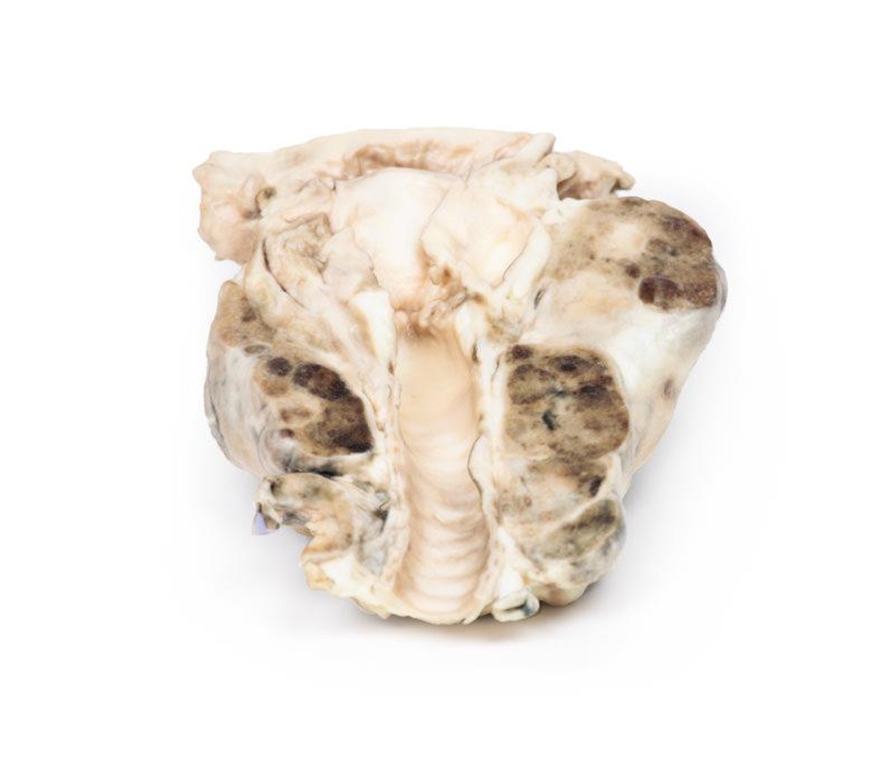
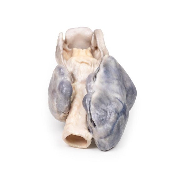
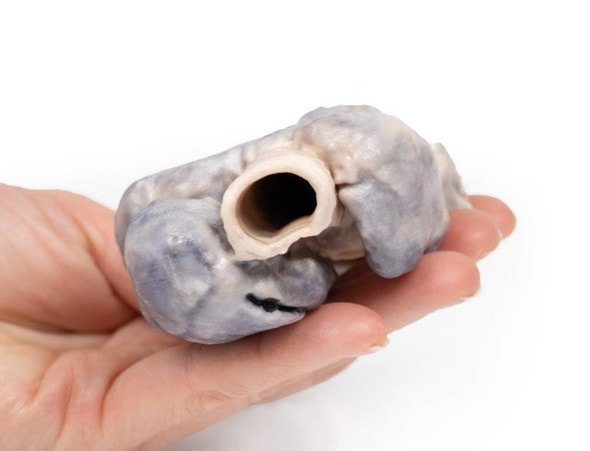
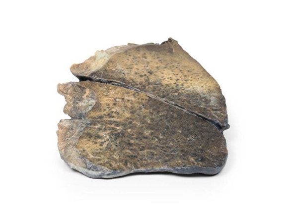
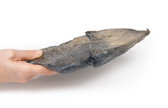
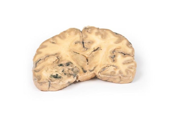
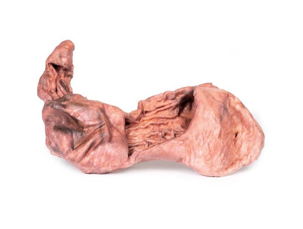
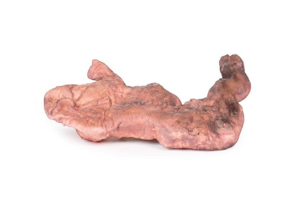
The specimen, removed at post-mortem, includes the base of the tongue, larynx and trachea. It has been cut in the coronal plane to allow a view of the internal laryngeal and tracheal anatomy. The thyroid gland is grossly enlarged particularly the right lobe, which extends superiorly and inferiorly, well beyond its normal margins when viewed from the anterior aspect. The cut posterior surfaces display many hyper- and hypopigmented nodules as well as cystic areas in both lobes. The tongue base, larynx and trachea appear relatively normal.
Further Information
Nodular goiter is most often detected simply as a mass or swelling in the neck but depending on size and location of growth may produce pressure symptoms on the trachea and the oesophagus. There may be difficulty in breathing, dysphagia, cough, and hoarseness. Paralysis of the recurrent laryngeal nerve may occur by an expanding goiter, but this is rare. Symptoms suggesting obstruction of the trachea including cough, stridor and shortness of breath may occur. Occasionally tenderness and a sudden increase in goiter size arise due to cystic expansion or hemorrhage into a nodule[1].
Causes of goiter include autoimmune disease (Hashimoto's thyroiditis, Grave's disease), the formation of one or more thyroid nodules and iodine deficiency. Goiter occurs when there is reduced thyroid hormone synthesis secondary to biosynthetic defects and/or iodine deficiency, leading to increased thyroid stimulating hormone (TSH). This stimulates thyroid growth as a compensatory mechanism to overcome the decreased hormone synthesis. Elevated TSH is also thought to contribute to an enlarged thyroid in the goitrous form of Hashimoto thyroiditis in combination with fibrosis secondary to the autoimmune process in this condition. In Grave's disease, the goiter results mainly from stimulation by the TSH receptor antibody[1].
Reference
1. Hughes et al. (2012) Goiter: Causes, investigation and management. Aust Family Physician, 41, 572-576.
Advantages of 3D Printed Anatomical Models
- 3D printed anatomical models are the most anatomically accurate examples of human anatomy because they are based on real human specimens.
- Avoid the ethical complications and complex handling, storage, and documentation requirements with 3D printed models when compared to human cadaveric specimens.
- 3D printed anatomy models are far less expensive than real human cadaveric specimens.
- Reproducibility and consistency allow for standardization of education and faster availability of models when you need them.
- Customization options are available for specific applications or educational needs. Enlargement, highlighting of specific anatomical structures, cutaway views, and more are just some of the customizations available.
Disadvantages of Human Cadavers
- Access to cadavers can be problematic and ethical complications are hard to avoid. Many countries cannot access cadavers for cultural and religious reasons.
- Human cadavers are costly to procure and require expensive storage facilities and dedicated staff to maintain them. Maintenance of the facility alone is costly.
- The cost to develop a cadaver lab or plastination technique is extremely high. Those funds could purchase hundreds of easy to handle, realistic 3D printed anatomical replicas.
- Wet specimens cannot be used in uncertified labs. Certification is expensive and time-consuming.
- Exposure to preservation fluids and chemicals is known to cause long-term health problems for lab workers and students. 3D printed anatomical replicas are safe to handle without any special equipment.
- Lack of reuse and reproducibility. If a dissection mistake is made, a new specimen has to be used and students have to start all over again.
Disadvantages of Plastinated Specimens
- Like real human cadaveric specimens, plastinated models are extremely expensive.
- Plastinated specimens still require real human samples and pose the same ethical issues as real human cadavers.
- The plastination process is extensive and takes months or longer to complete. 3D printed human anatomical models are available in a fraction of the time.
- Plastinated models, like human cadavers, are one of a kind and can only showcase one presentation of human anatomy.
Advanced 3D Printing Techniques for Superior Results
- Vibrant color offering with 10 million colors
- UV-curable inkjet printing
- High quality 3D printing that can create products that are delicate, extremely precise, and incredibly realistic
- To improve durability of fragile, thin, and delicate arteries, veins or vessels, a clear support material is printed in key areas. This makes the models robust so they can be handled by students easily.

