Description
Realistic Imaging Practice for Veterinary Professionals
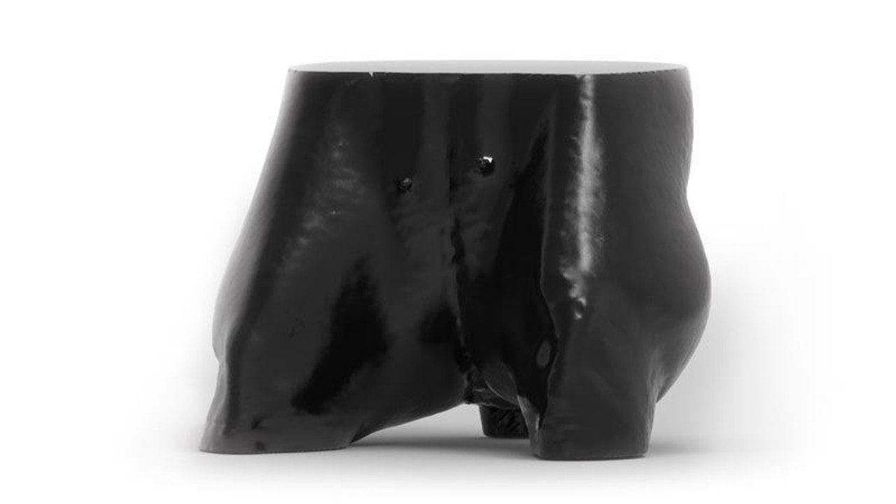
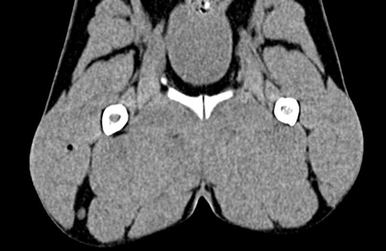
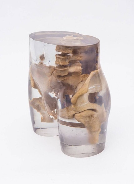
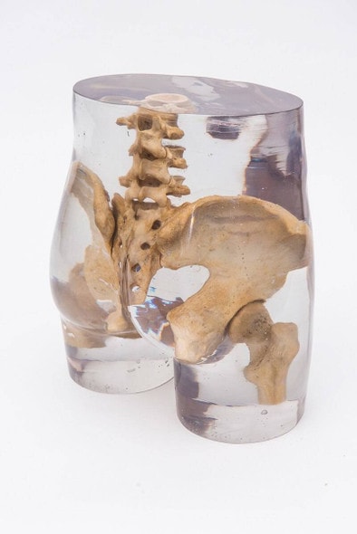
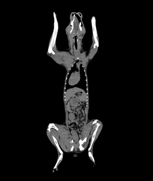
Train veterinary students and radiologic technologists with precision using the Dog Pelvis Phantom for CT and X-Ray. This anatomically accurate model replicates the bony structure of a canine pelvis, making it ideal for imaging practice and diagnostic training. Built from realistic materials, it offers lifelike radiodensity under both CT and X-ray imaging. Enhance your training curriculum today with this essential educational tool.
Master Canine Imaging Techniques with Confidence
This phantom is specifically designed to simulate real canine anatomy, enabling learners to practice and refine their imaging techniques in a controlled, repeatable environment. It supports essential learning for orthopedic evaluations, pelvic trauma assessments, and anatomical orientation. Ideal for veterinary schools, radiology departments, and animal hospitals, this model boosts clinical confidence before live patient interaction.
Train Like It’s Real—Because It Practically Is
- Provides highly detailed pelvic bone structures for accurate diagnostic simulation - Perfect for X-ray and CT imaging of canine pelvic anatomy - Great for evaluating equipment settings, exposure techniques, and positioning - Supports advanced veterinary imaging education for students and professionals - Trusted by veterinary training programs and imaging facilities worldwide
Key Features of the Dog Pelvis Phantom
- Realistic bone density and anatomical accuracy - Radiopaque material suitable for both CT and X-ray imaging - Durable, long-lasting construction for repeated use - Easy to handle and transport
Technical Specifications of the Product
- Product weight: 11 lbs.
- Included with purchase:
- 1 x Dog Pelvis Phantom
- 1 x Product Manual
- 1 x Product Case

