- Home
- 3D Printed Models
- Animal Prints
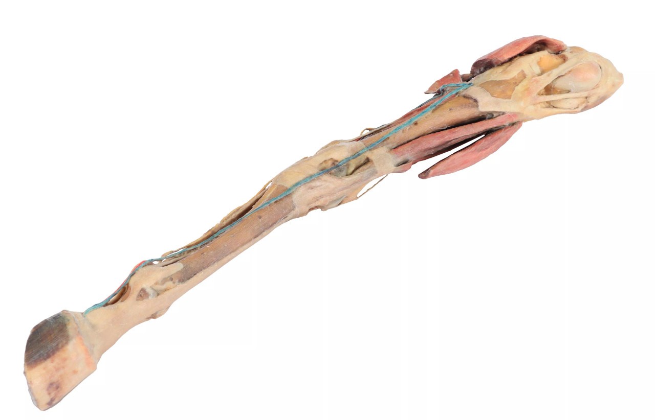
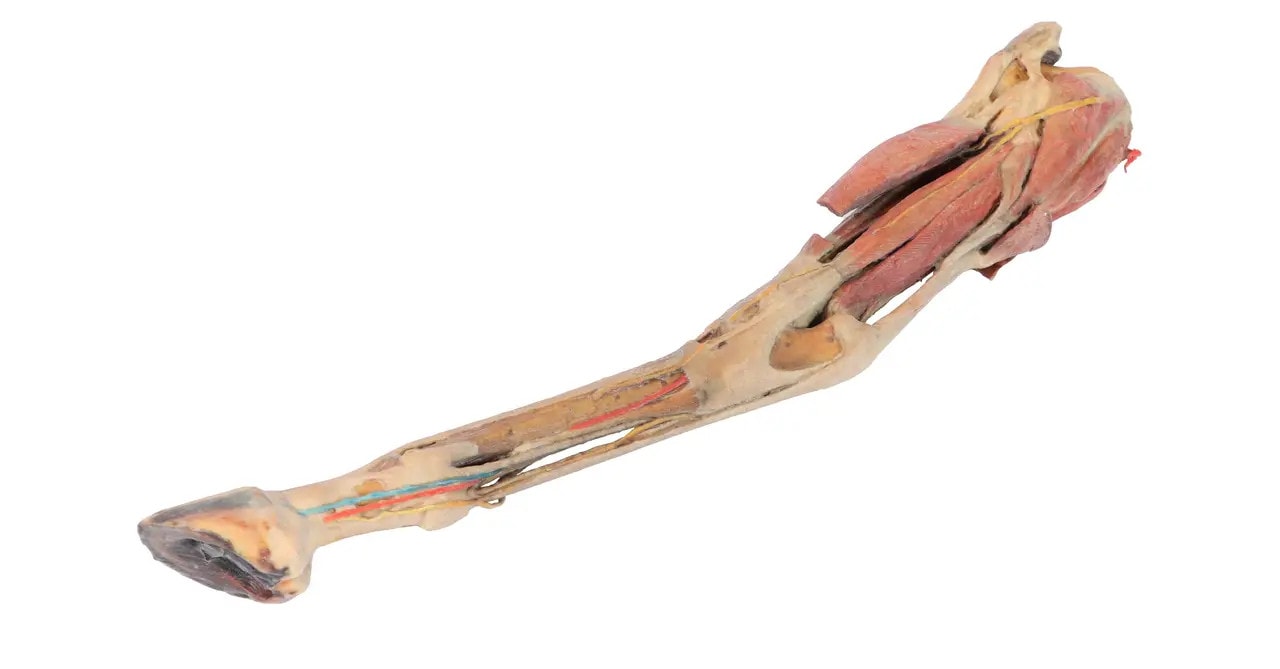
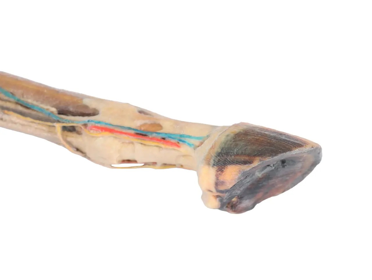
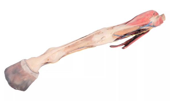
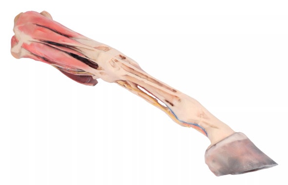
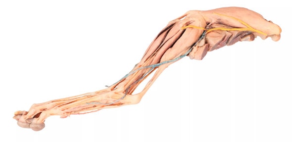
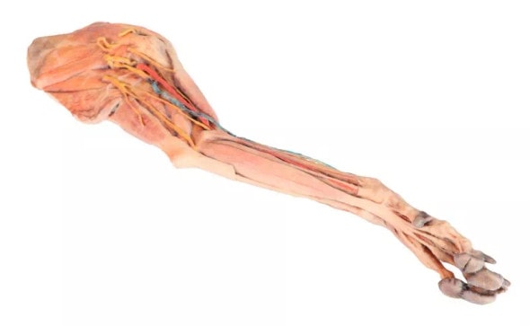
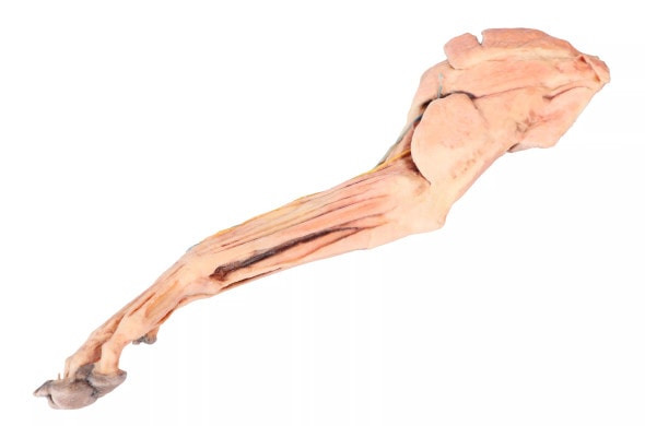
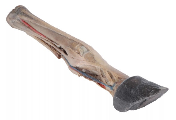
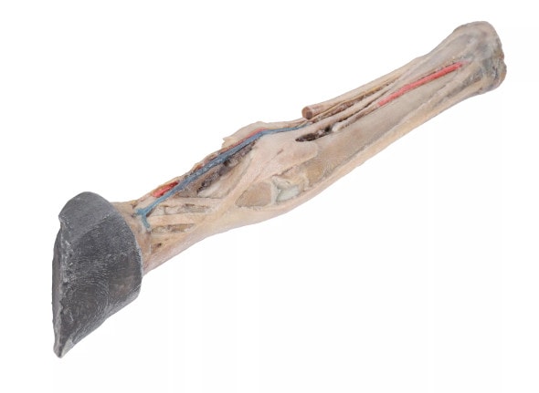
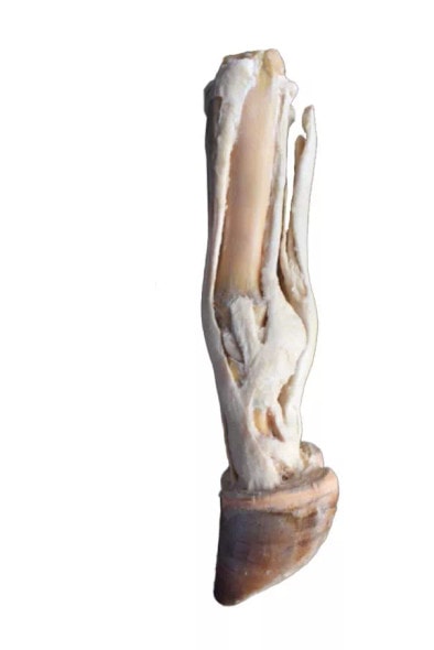
- 3D Printed 1/3 Size Horse Hindlimb - Muscles, Tendons, Ligaments, Vessels, and Nerves Distal to the Stifle
Description
Perfectly Scaled Detailed Equine Hindlimb Anatomy
This 1/3 scale dissection 3D Printed model of a horse's hindlimb offers an accurate and detailed view of the muscles, tendons, ligaments, vessels, and nerves located distal to the stifle joint. Crafted from real anatomical specimens, it is an indispensable tool for understanding the complex structure of the equine lower limb. Compact yet highly informative, the model is ideal for both classroom and clinical use. Add it to your veterinary teaching collection or equine practice today,
Precision Teaching Tool for Veterinary and Farrier Training
Designed for in-depth anatomical education, this model is particularly useful for learning equine locomotion biomechanics, neurovascular structures, and musculoskeletal function. It allows veterinary students, farriers, and equine therapists to explore the functional anatomy of the distal hindlimb in a hands-on format. The model is also beneficial in diagnosing and explaining common issues such as suspensory injuries, tendonitis, or circulatory problems in horses. Whether used for instruction or client education, it delivers high educational value.
See What Lies Beneath: A Must-Have for Equine Professionals
- Excellent for Various Learning Environments: Anatomy Labs, Veterinary Courses, or Farrier Training
- Provides Clear Visuals: Distal Limb Components for injury explanation and prevention
- Encourages Hands-on Learning: more effective retention of complex anatomy
- Supports Professional Diagnosis: helps professionals in diagnosis in and communicating equine limb disorders.
- Compact Size: ideal for mobile education or demonstration settings
Product Features at a Glance
- 1/3 Life Size Dissection of Horse Hindlimb (distal to stifle)
- Muscles, Ligaments, Tendons, Vessels, and Nerves in high Detail
- Real Anatomical Specimen with Lifelike accuracy
- Durable and Designed for Repeated Educational Use
Technical Specifications of the Product
- Product dimensions: 7.9 in x 7.9 in x 11.8 in.
- Product weight: 2.5 lbs
- Included with purchase
- 1 x Muscles, Tendons, Ligaments, Vessels, and Nerves Distal to the Stifle - Horse Hindlimb 3D Printed 1/3 Size Model
- 1 x Product Manual


