Description
Uncover the Structure Beneath the Hoof
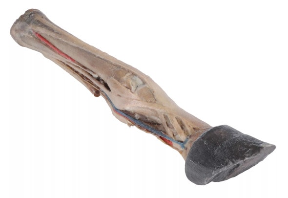
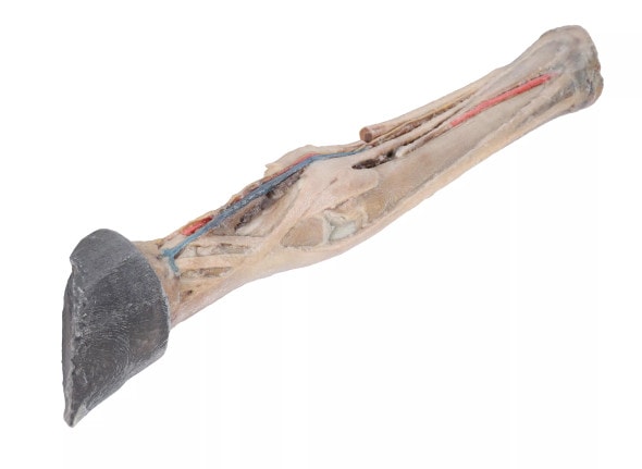
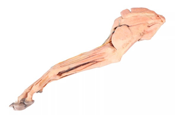
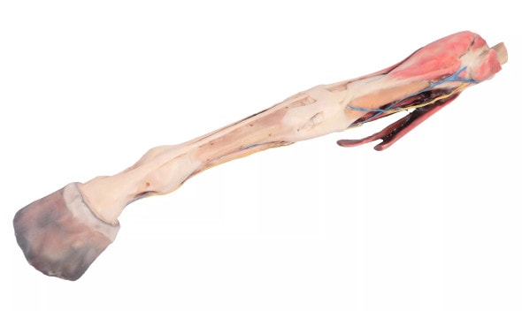
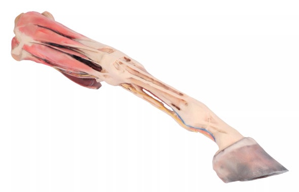
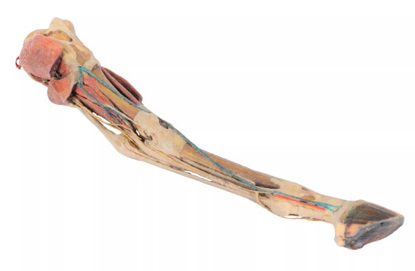
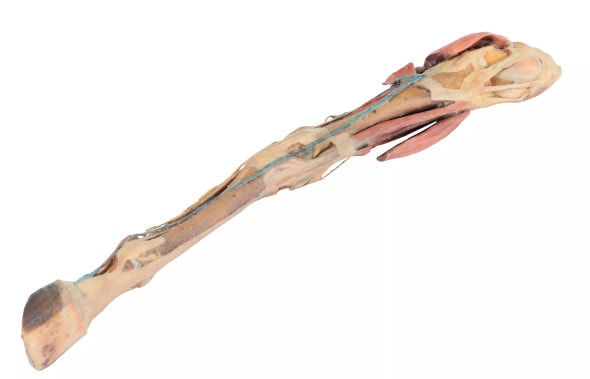
This high-quality 3D Printed anatomical model of the ox foot displays detailed dissections of tendons and ligaments, offering a clear view of essential musculoskeletal structures. Based on real specimens, it provides an authentic and accurate representation of bovine foot anatomy. Ideal for veterinary educators, students, and livestock professionals, this model helps bring textbook knowledge into three-dimensional reality. Add this essential teaching tool to your anatomical collection today.
Essential for Veterinary and Bovine Education
This model is designed to support the study of bovine anatomy, particularly in understanding the structural and functional role of tendons and ligaments in the distal limb. It's perfect for veterinary schools, anatomy labs, and continuing education in large animal care. Users can explore topics such as locomotion, hoof health, and lameness diagnosis in a tactile, engaging way. It's especially helpful in training future veterinarians and hoof trimmers in proper limb care.
Why Every Bovine Educator Needs This Model
- Offers a realistic look at the internal anatomy of the ox foot
- Enhances student engagement and retention with hands-on learning
- Aids in explaining hoof pathologies and treatment to clients or students
- Compact and durable -- Ideal for classroom and mobile teaching
- Suitable for both beginners and advanced learners in veterinary science
Detailed Features at a Glance
- Real-specimen-based ox foot dissection
- Shows flexor and extensor tendons, ligaments, and joint structures
- Mounted securely for classroom use of demonstrations
- Crafted with durable materials for long-term handling
- Accurately scaled for teaching and clinical reference
Technical Specifications of the Product
- Product dimensions: 7.9 in x 7.9 in x 7.9 in.
- Product weight: 2.5 lbs.
- Included with purchase:
- 1 x Ox foot - tendons and ligaments 3D Printed Model
- 1 x Product Manual


