- Home
- Medical Task Trainers
- Medical Imaging Task Trainers
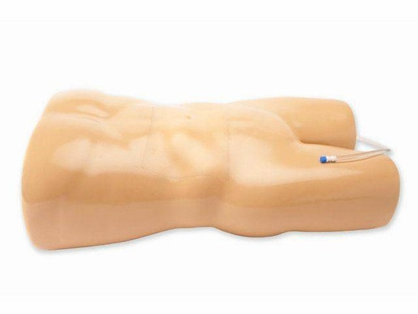
- Transthoracic Echocardiography and Pericardiocentesis Ultrasound Training Model
Description
See & Treat: Real-Time Echo with Life-Saving Pericardiocentesis Training
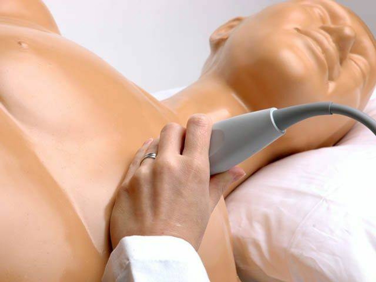
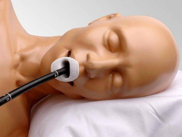
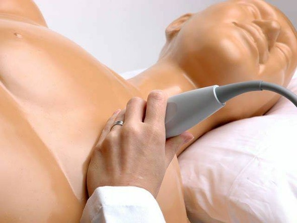
Take your cardiac ultrasound training to the next level with the Transthoracic Echocardiography and Pericardiocentesis Ultrasound Training Model. This comprehensive simulator combines realistic heart imaging with hands‑on pericardial access practice for a powerful, dual‑purpose learning experience. Its lifelike anatomy challenges learners to identify effusions and perform pericardiocentesis under ultrasound guidance. Empower your training program today—start mastering these critical cardiac procedures!
Targeted Training for Cardiac Diagnostic and Interventional Skills
Designed for clinicians refining transthoracic echo (TTE) and emergency pericardiocentesis, this model supports both diagnostic imaging and procedural proficiency. Trainees can practice acquiring standard echo windows, interpreting fluid collections, and inserting needles safely into the pericardial space. Ideal for cardiologists, ER physicians, anesthesiologists, critical care teams, and echo techs, this model builds essential cardiac skills in one integrated platform.
From Simulation Lab to Critical Care Command
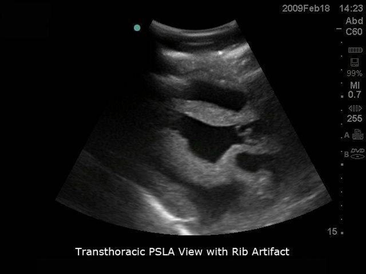
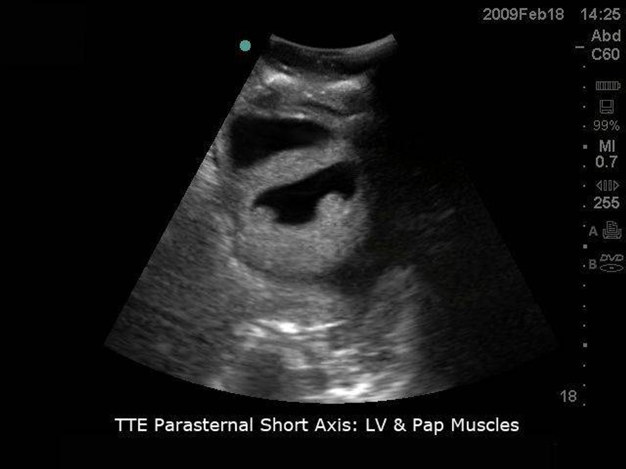
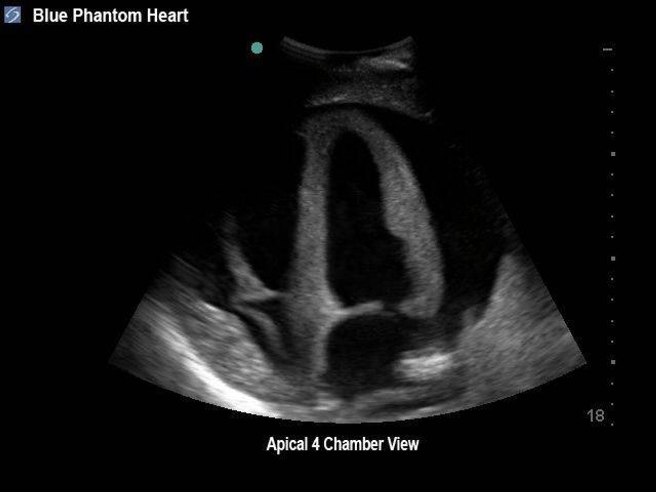
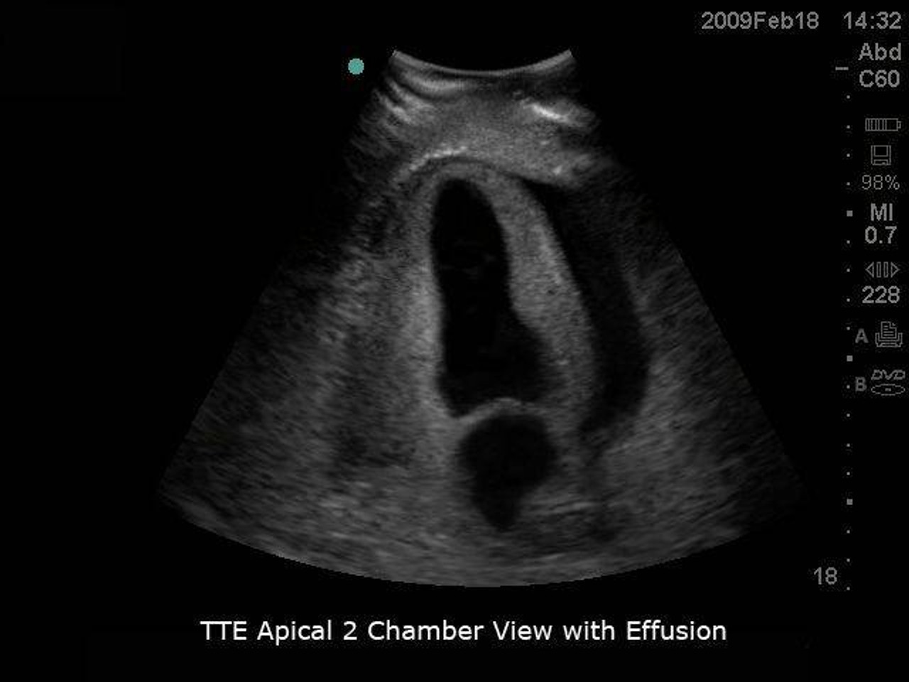
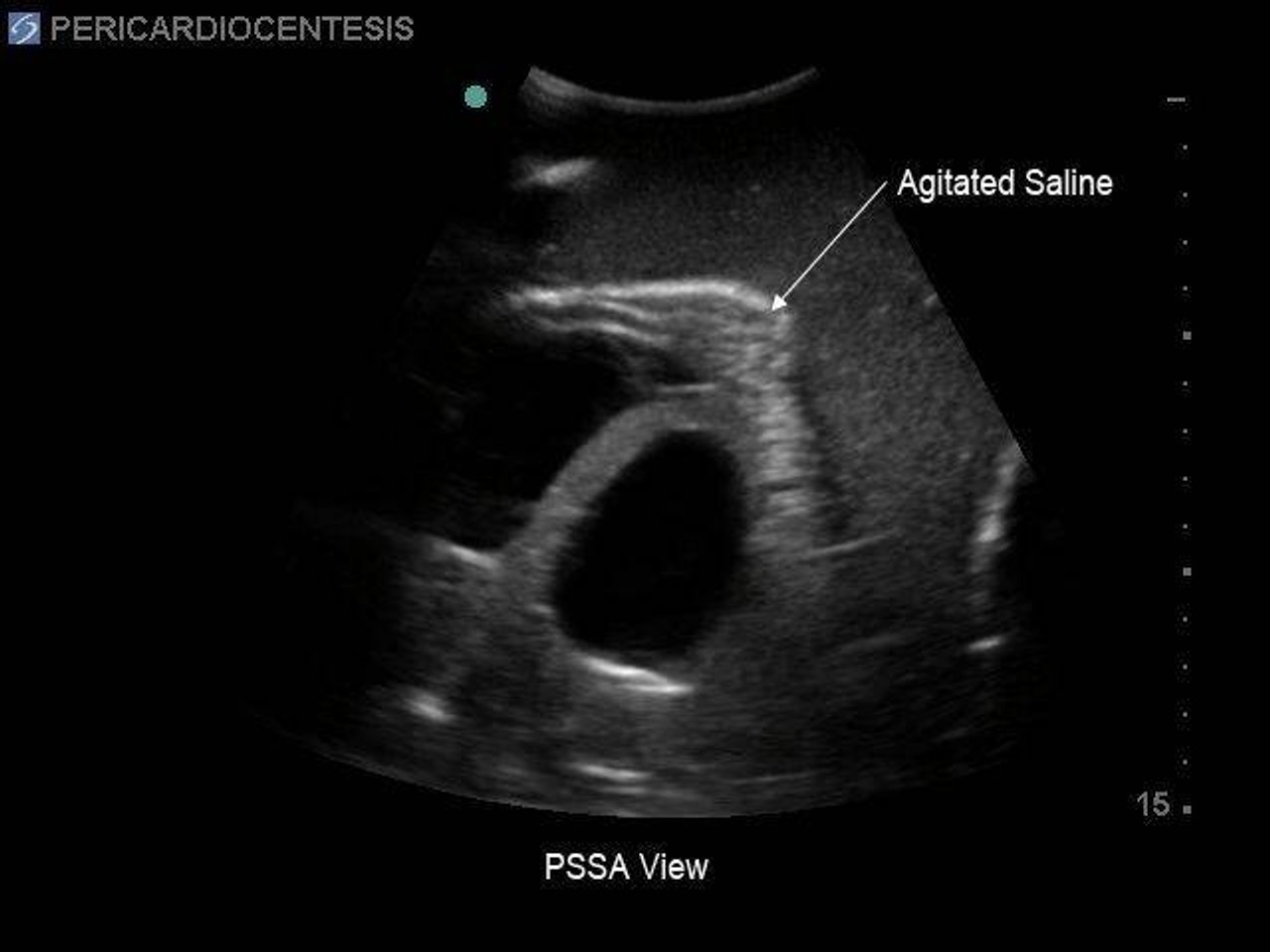
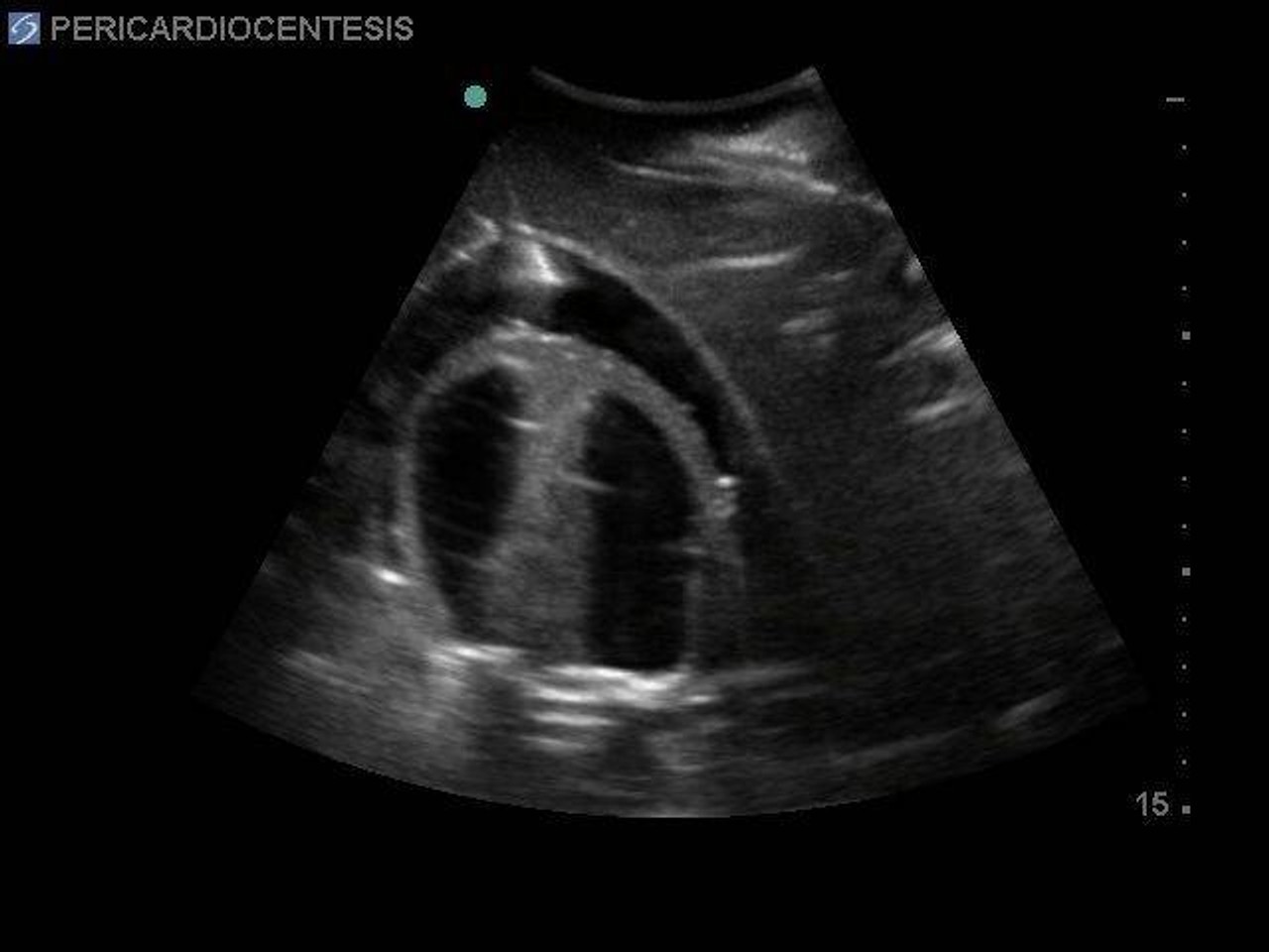
- Experience lifelike echocardiographic views, including apical and parasternal windows, for confident image acquisition
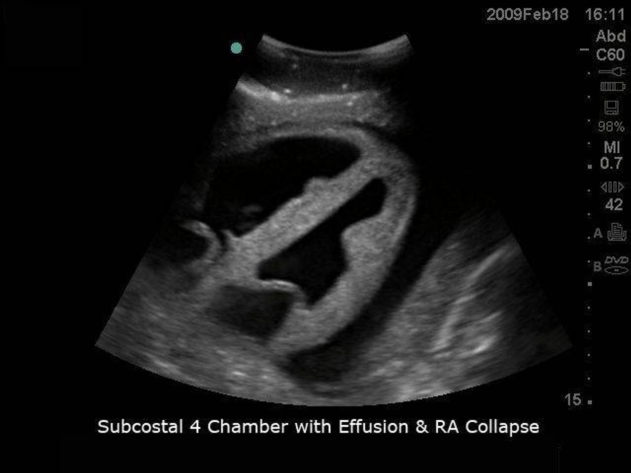
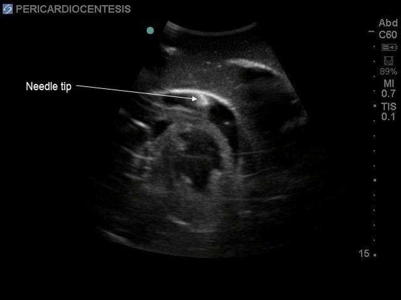
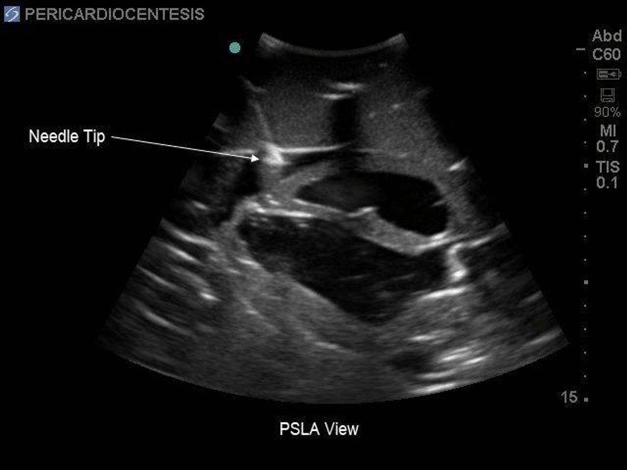
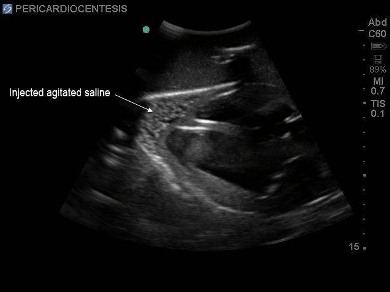
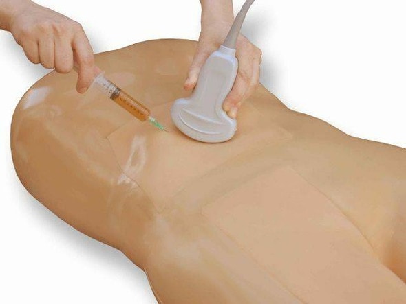
- Balance image interpretation with procedural action by detecting pericardial effusions and aspirating fluid precisely
- Ideal for emergency, ICU, cardiology, and anesthesia training programs, incorporating real‑time needle guidance with ultrasound
- Enhances learner competence and confidence in diagnosing tamponade and performing pericardiocentesis
- Durable and reusable model supports repeatable practice for both novice and experienced providers
Features That Make Cardiac Training Exceptional
- High-fidelity simulated cardiac anatomy compatible with ultrasound imaging
- Functional pericardial cavity with realistic fluid pressure and needle feedback
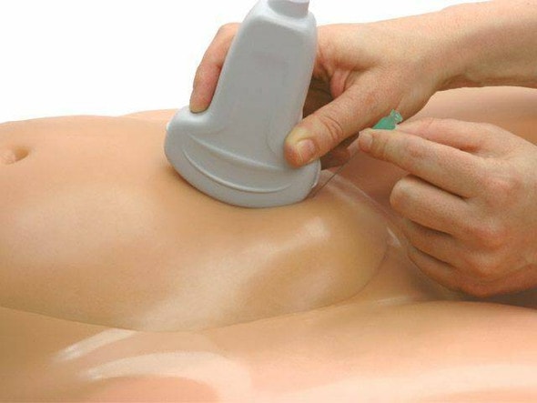
- Supports in-plane and out-of-plane needle approaches under ultrasound guidance
- Compact size and durable materials built to last through frequent simulation sessions
- Compatible with most standard transthoracic ultrasound probes
Technical Specifications of the Product
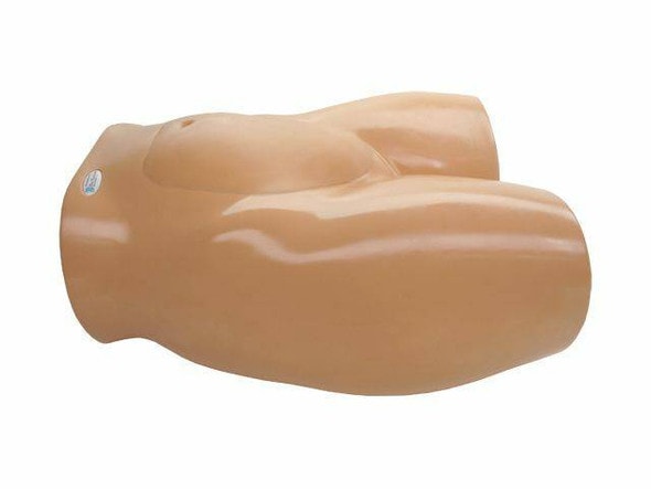
- Product dimensions: 15 in x 21 in x 40 in.
- Product weight: 110 lbs.
- Included with purchase:
- 1 × Transthoracic Echo & Pericardiocentesis Training Model
- 1 × Product Manual
- 1 × Fluid Refilling Kit

