Description
Unlock the Complexity of Equine Locomotion
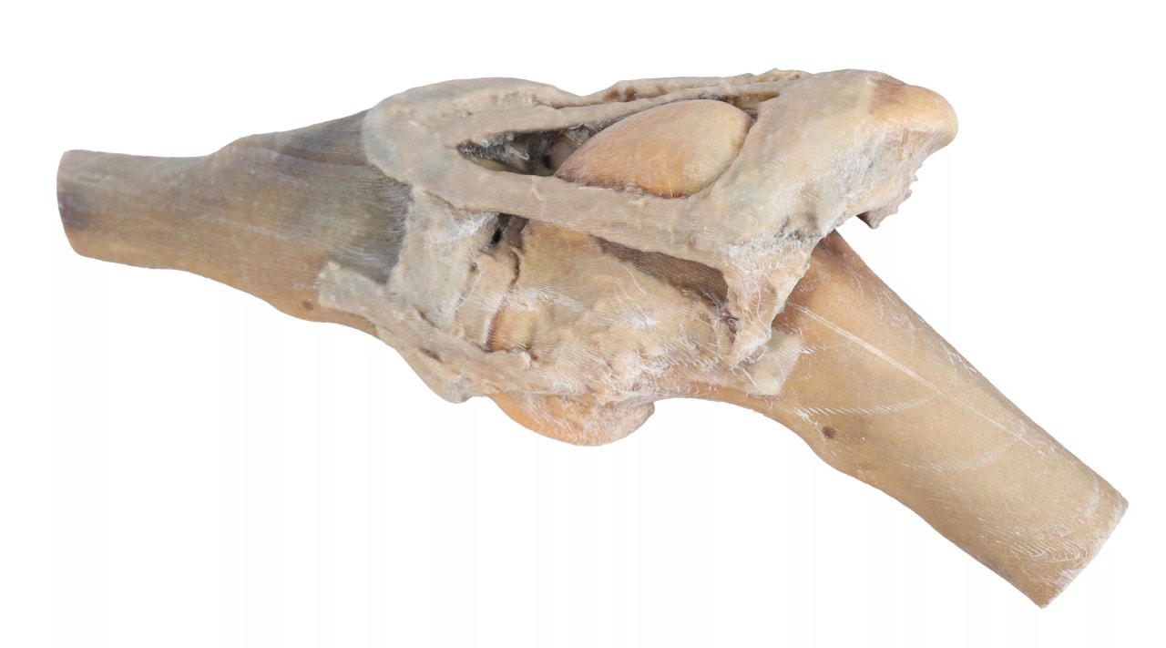
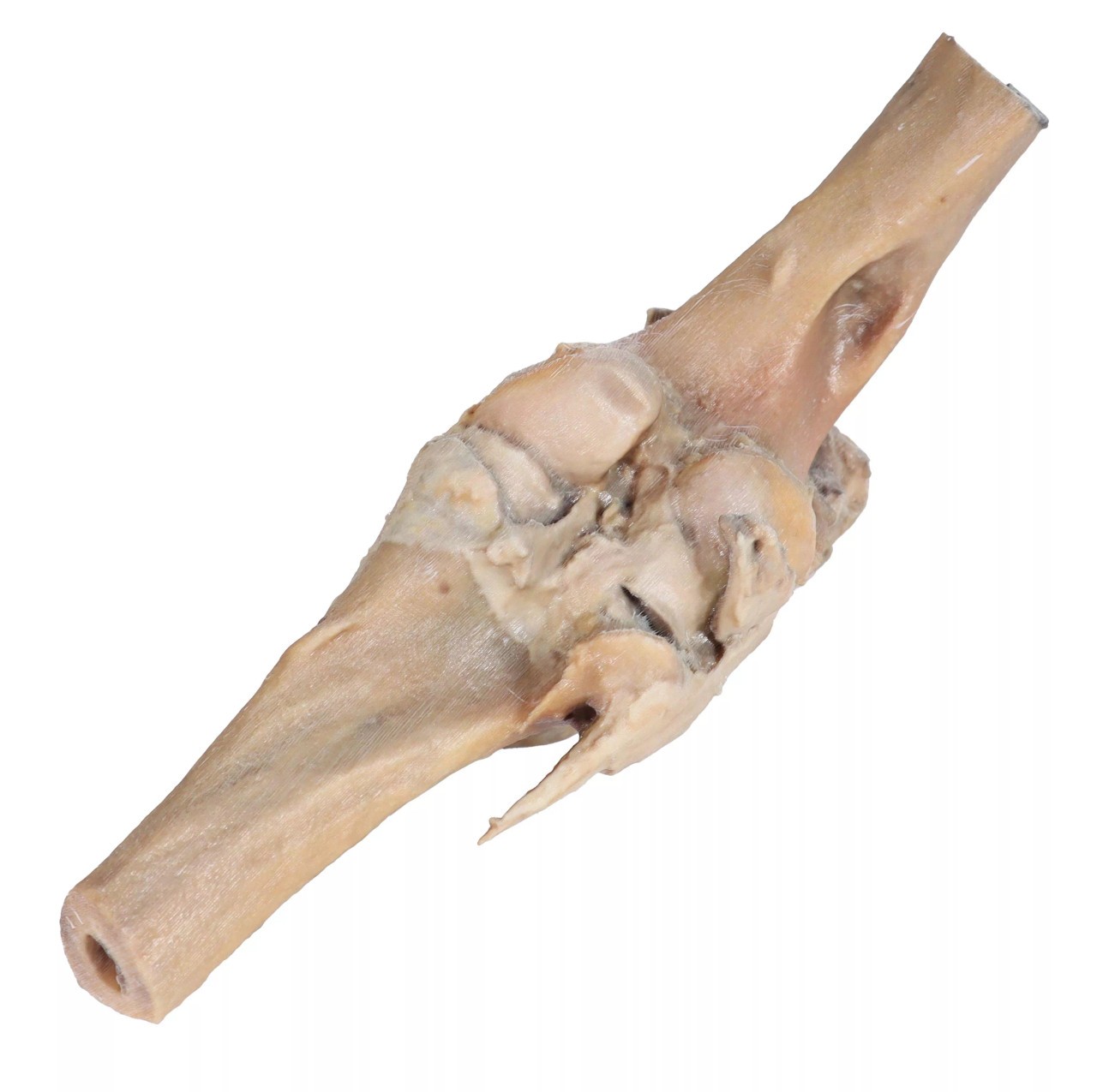
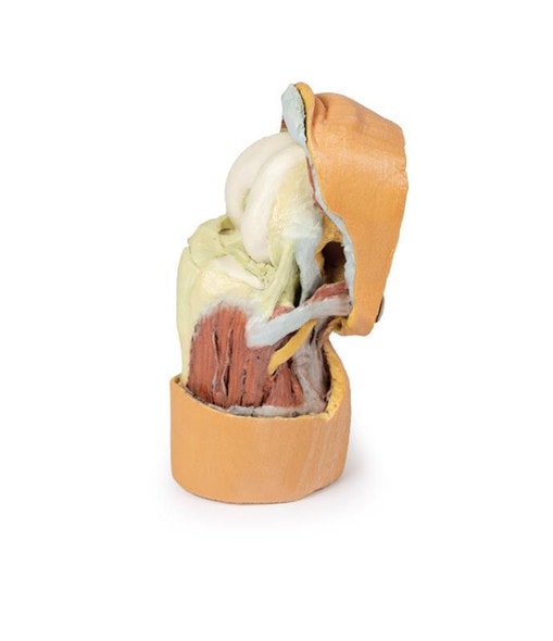
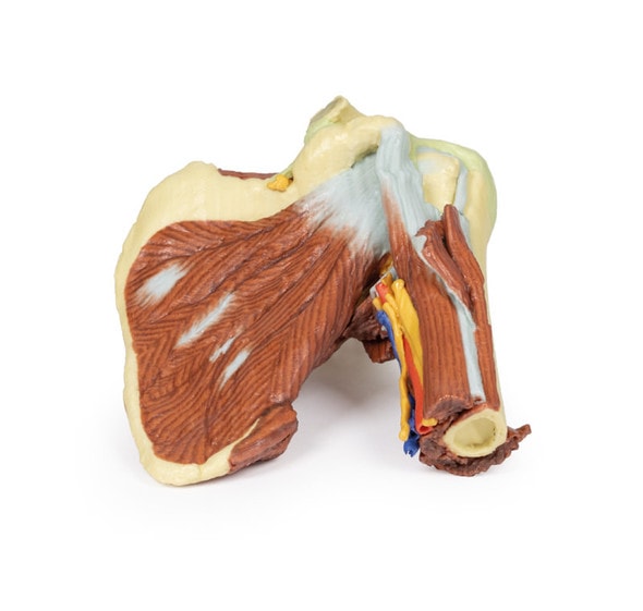
The 3D printed Horse Stifle Joint Dissection Model offers a realistic and detailed view of the stifle joint's internal anatomy. Carefully dissected to show ligaments, tendons, joint surfaces, and Cartlidge, this model is perfect for advanced study and professional reference. Veterinary educators, students, and equine specialists can use it to reinforce anatomical understanding, aid surgical planning, and improve client communication. Add this essential tool to your anatomical collection to enhance equine or orthopedic training.
Designed for Teaching, Trusted for Practice
This model provides a hands-on resource for exploring the biomechanics and structural relationships of the horse stifle joint. Users can study the patella, femur, tibia, and associated connective tissues to better understand injury mechanisms and rehabilitation. Ideal for courses in equine orthopedics, this model is also a great tool for veterinary practitioners when explaining conditions or procedures related to stifle joint dysfunction.
Key Benefits
- Enhances Practical Learning - of horse joint anatomy and mechanics
- Excellent Visual Aid - for veterinary education in clinical settings
- Realistic Representation - improves understanding of complex ligament structures
- Designed for Use in Classrooms
- Supports Planning - surgical planning and injury assessment for equine practitioners
Key Features
- Detailed Dissection - shows internal structures of equine stifle joint
- 3D Printed - modeled on real anatomical specimens
- Color Coded Elements - for easy identification of tissues
- Durable - suited for repeated use in a variety of settings
Technical Specifications of the Product
- Product dimensions: 7.9 in x 7.9 in x 11.8 in.
- Product weight: 2.5 lbs
- Included with purchase:
- 1x Horse Stifle Joint Dissection 3D Printed Model
- 1 x Product Manual


