Description
Explore the Foundations of Equine Structure with Precision
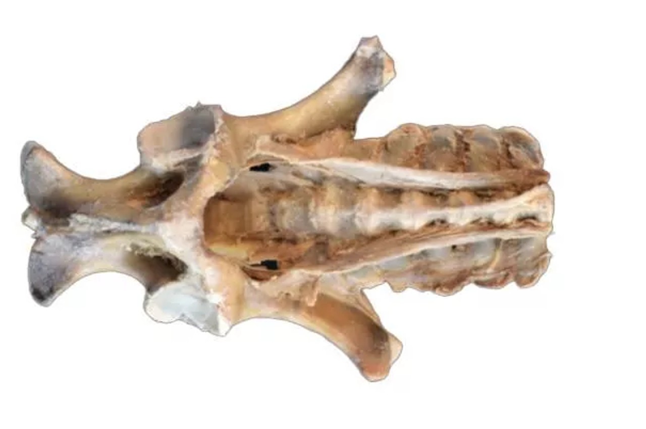
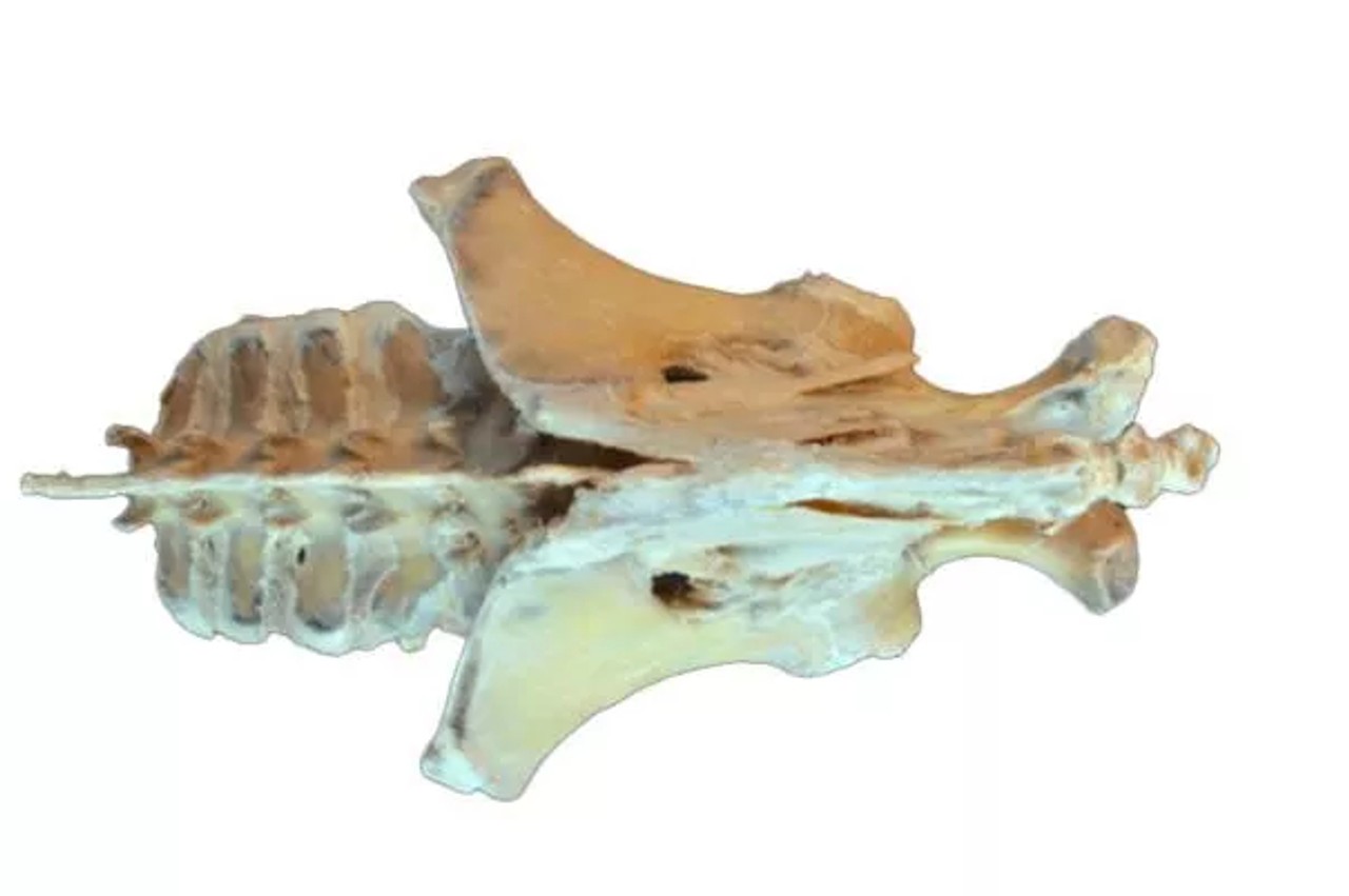
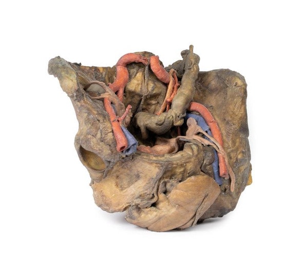
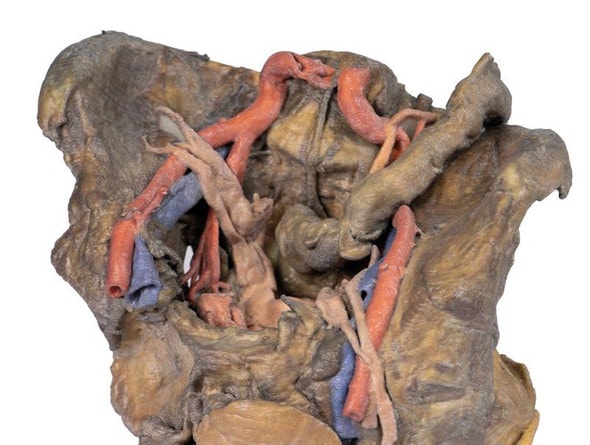
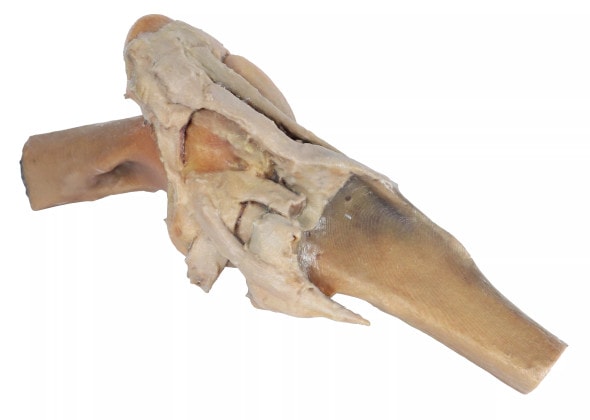
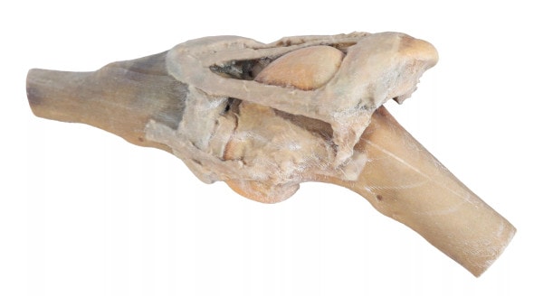
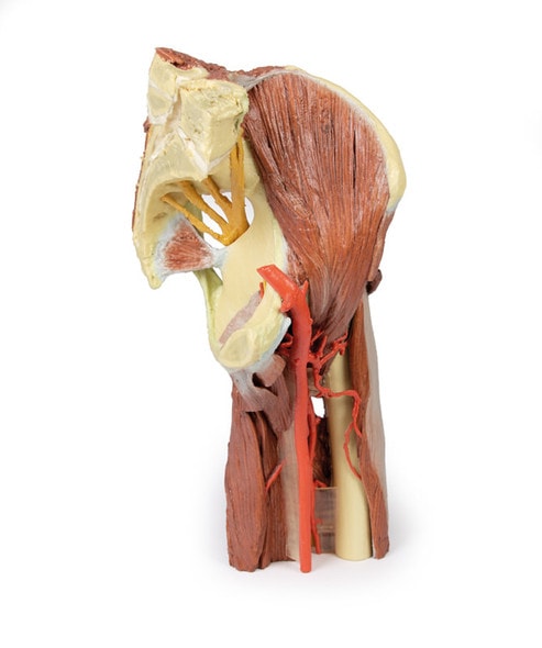
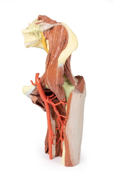
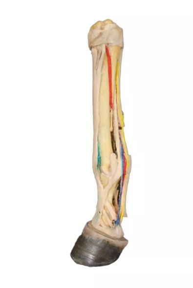
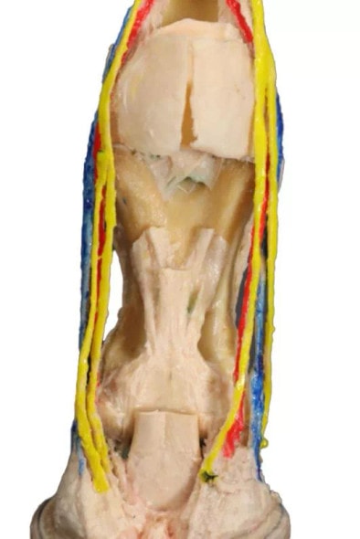
The 3D Printed Horse Foal Pelvis Dissection - Ligaments offers an anatomically accurate look at the young equine pelvic region, highlighting key ligamentous structures. This model is designed for veterinary professionals and educators seeking a detailed reference for the os coxae, sacroiliac joint, and associated connective tissues. Ideal for teaching equine development, orthopedic planning, or clinical explanation. Add this model to your anatomy lab or veterinary collection to elevate your instruction and understanding.
Built for Education, Designed for Clinical Relevance
This dissection model serves as a powerful educational aid for teaching musculoskeletal anatomy in developing horses. Veterinary students can use it to study bone-ligament relationships, while instructors can reinforce concepts like load-bearing anatomy, pelvic symmetry, and foal development. Clinicians may find it useful for diagnosing or explaining developmental issues in young horses. It supports hands-on learning for equine orthopedics and biomechanics.
Understand Growth, Movement, and Structure in Foals
- Built for Education - perfect for veterinary schools, equine specialists, and anatomy educators
- Anatomical Clarity - offers a clear view of the pelvis, sacrum, and major ligaments in a foal specimen
- Hands on Instruction - Supports teaching about equine musculoskeletal development
- Enhances Learning - about developmental orthopedic conditions
Key Features of the Model
- Realistic Dissection - horse foal pelvis including visible ligaments
- Anatomically Accurate - derived from actual specimens
- Color Coded Structures - for easier learning and identification
- Ideal for Display - compact, easy to use in teaching enviroments
Technical Specifications of the Product
- Product dimensions: 7.9 in x 7.9 in x 11.8 in.
- Product weight: 2.5 lbs
- Included with purchase:
- 1 x Horse (foal) Pelvis Dissection - Ligaments 3D Printed Model
- 1 x Product Manual

