Description
Dive Deep Into Equine Foot Anatomy
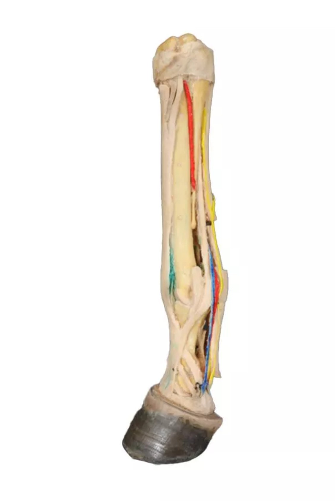
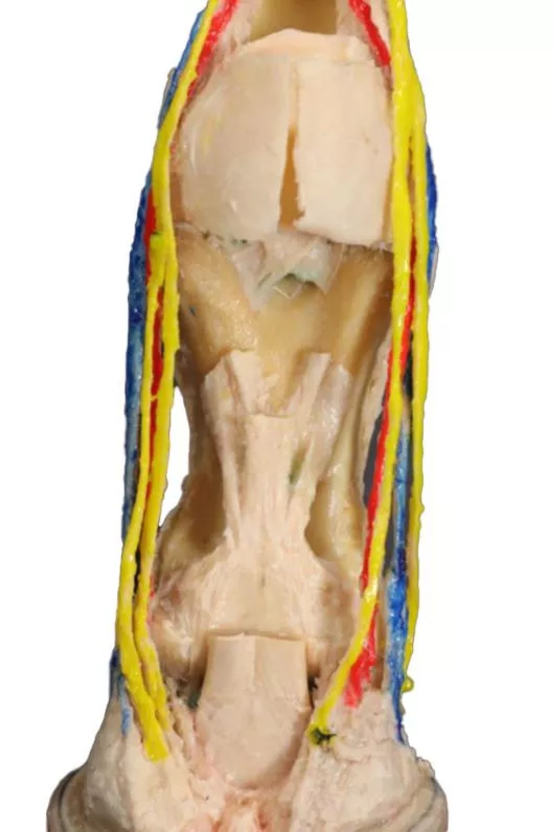
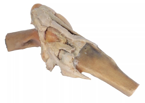
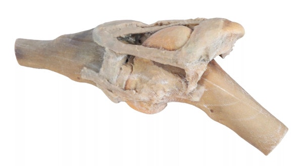
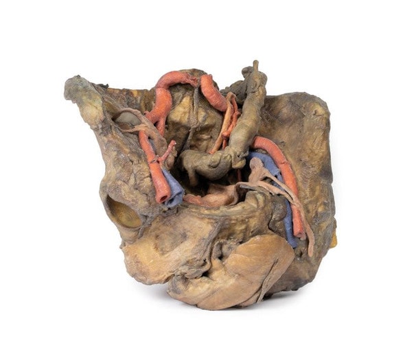
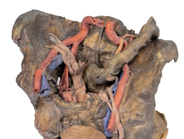
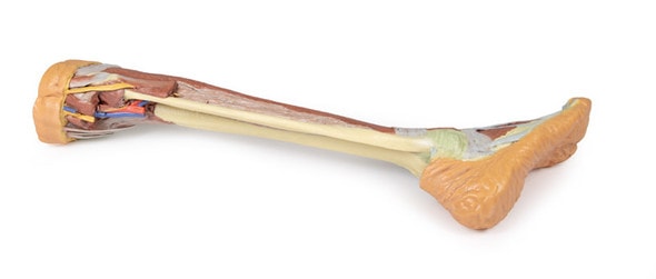
Uncover the complexity of the equine distal limb with the Deep Palmar - Horse Foot Dissection 3D Printed Model. This anatomically accurate teaching tool highlights the intricate structures of the deep palmar region, including flexor tendons, ligaments, and neurovascular components. Designed from real anatomical specimens, it delivers clarity and realism for advanced anatomy education. Enhance your training environment or clinical explanations with this essential equine model
Designed for Advanced Learning and Clinical Insight
This model provides and in-depth look at the deep palmar aspect of the horse foot, ideal for studying equine orthopedic health, lameness evaluation, or foot biomechanics. Veterinary students and professionals can gain practical insight into the digital flexor apparatus and related vasculature. It's also a valuable reference for farriers and equine therapists to better understand internal hoof anatomy. The model supports targeted instruction in equine musculoskeletal and neurological systems.
Where Precision Meets Practicality in Hoof Education
- Perfect for Veterinary Educators, Farriers, and Equine Orthopedic Specialists
- Highlights the deep digital flexor tendon, suspensory ligament, and palmar neurovascular structures
- Supports diagnostic discussions about navicular syndrome, deep tendon injuries, or vascular compromise
- Enhances hands-on learning for hoof care and lameness evaluation
- Valuable for client education or anatomy lab demonstrations
Key Features of the Model
- Realistic dissection of the equine deep palmar region
- Cleary shows the tendons, ligaments, and nerve-vessels structures
- Based off real anatomical specimens for maximum accuracy
- Compact and Durable - Ideal for mobile teaching or clinical use
Technical Specifications of the Product
- Product dimensions: 7.9 in x 7.9 in x 11.8 in
- Product weight: 2.5 lbs
- Included with purchase:
- 1 x Deep Palmar Dissection - Horse Foot 3D Printed Model
- 1 x Product Manual

