- Home
- Anatomy Models
- Organ Models
- Reproductive Organ Models
- Pregnancy Models
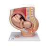
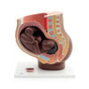
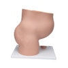
- Pregnancy Pelvis Model with 40-Week Fetus, 3-Parts





Description
Female Pelvis Median Section Model
40th Week of Pregnancy with Removable Fetus
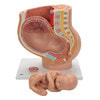
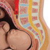
This detailed female pelvis model illustrates a median section of the pelvis during the 40th week of pregnancy. Designed for accurate anatomical study, the model clearly demonstrates the position of the fetus shortly before birth, making it ideal for medical education, midwifery training, and patient communication.
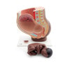
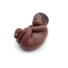
The removable fetus allows closer examination of its placement within the uterus and supports a better understanding of the birthing process and late-stage pregnancy anatomy.
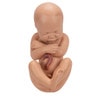
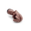
For developmental comparison, the model also includes an embryo in the 3rd month of pregnancy, mounted on the base. This additional feature helps visualize the changes in size and development throughout pregnancy.
Key Features
Detailed Median Section
The anatomical cross-section provides a clear view of the maternal pelvis and fetal position during late pregnancy, supporting realistic demonstration and study.
Removable Fetus
The fetus can be easily removed for enhanced observation and hands-on learning during lectures, training sessions, or patient explanation.
Embryo Comparison Model
Includes a 3rd month embryo model for direct comparison of prenatal development stages.
Designed for Educational Use
Suitable for use in:
- Universities and medical schools
- Midwifery and obstetrics training
- Clinical teaching environments
- Patient education and demonstration
Summary
A high-quality female pelvis anatomy model designed to demonstrate fetal positioning and prenatal development during the final stage of pregnancy — ideal for teaching, training, and clinical explanation.