- Home
- Promotions
- Product Kits, Bundles, & Sets
- Anatomy Model Kits, Bundles, and Sets
- The Senses Anatomy Model Set
Description
See, Hear, Touch, Taste, and Smell—All in One Stunning Model Set
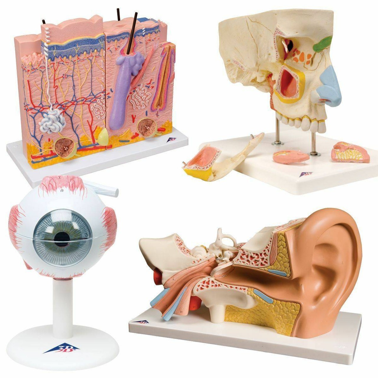
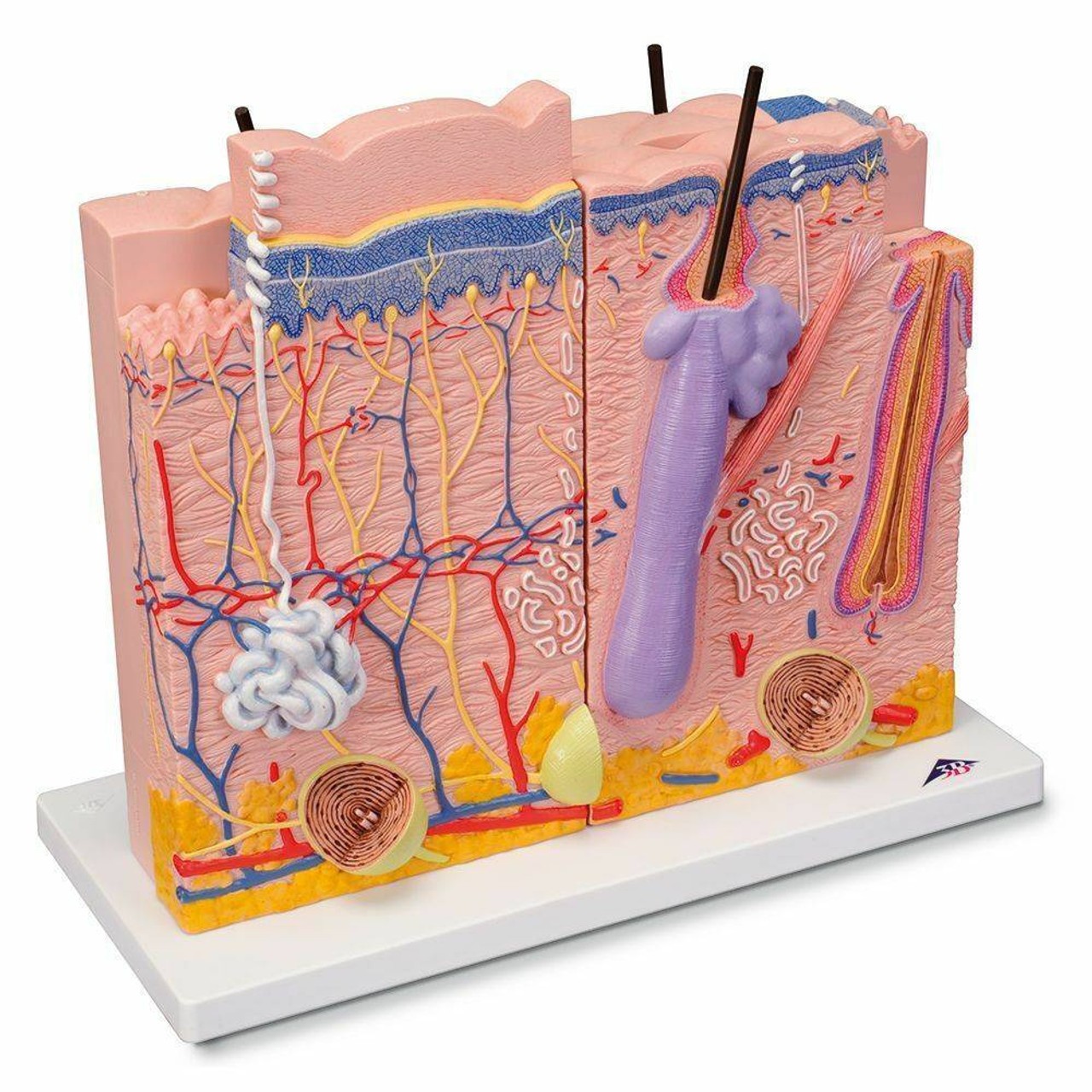
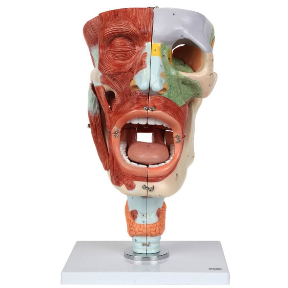
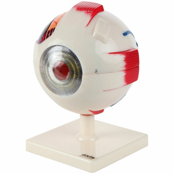
Engage your students in a complete sensory experience with The Senses Anatomy Model Set. This collection includes detailed representations of the five primary sense organs: eye, ear, skin, tongue, and nose—perfectly designed for clear, hands-on learning. Whether you're in the classroom or the clinic, this set simplifies complex anatomical concepts with colorful precision. Order now and give your learners a well-rounded understanding of human perception!
Teach the Five Senses with Five Powerful Tools
This anatomy model set is designed to provide a tactile and visual approach to understanding how humans perceive the world. Ideal for teaching biology, anatomy, or health science, it allows users to identify the key anatomical structures responsible for sight, hearing, touch, taste, and smell. Students can explore each organ system in isolation while appreciating the interconnectedness of the senses. It's a perfect fit for interactive demonstrations and anatomy-based curriculum.
Bring Human Perception to Life in the Lab or Classroom
Help students connect abstract theory to real-world biology:
- Includes 5 distinct models representing each of the five senses
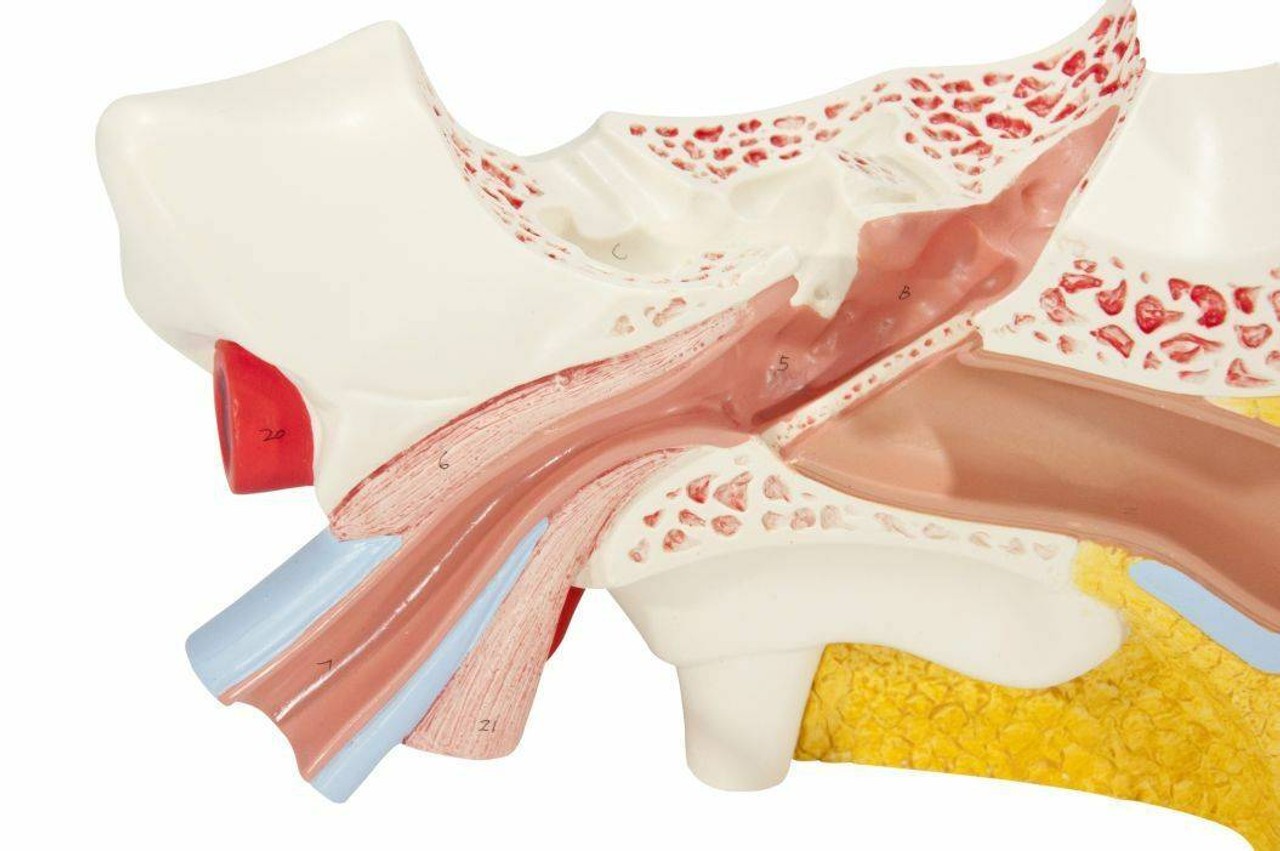
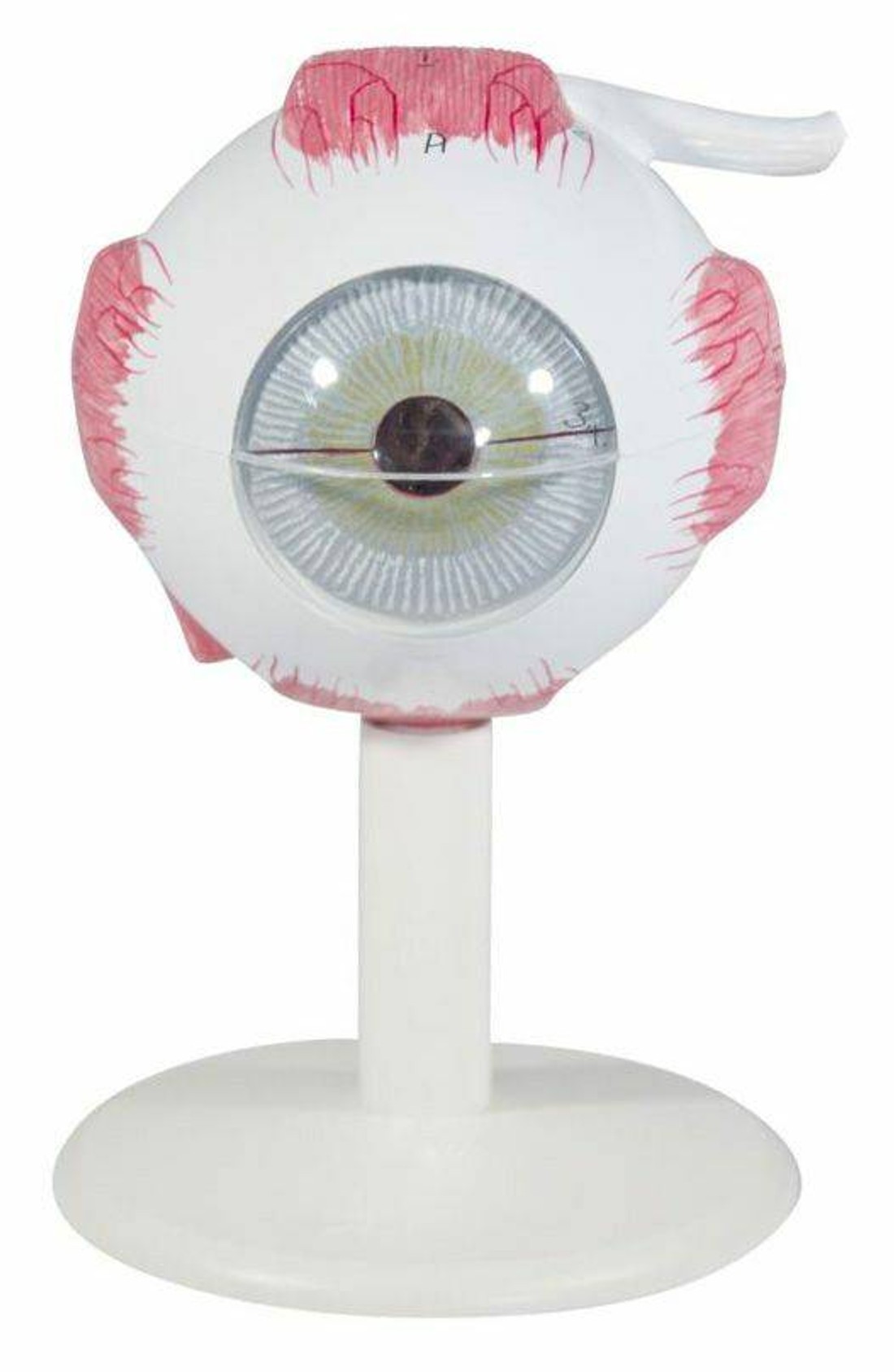
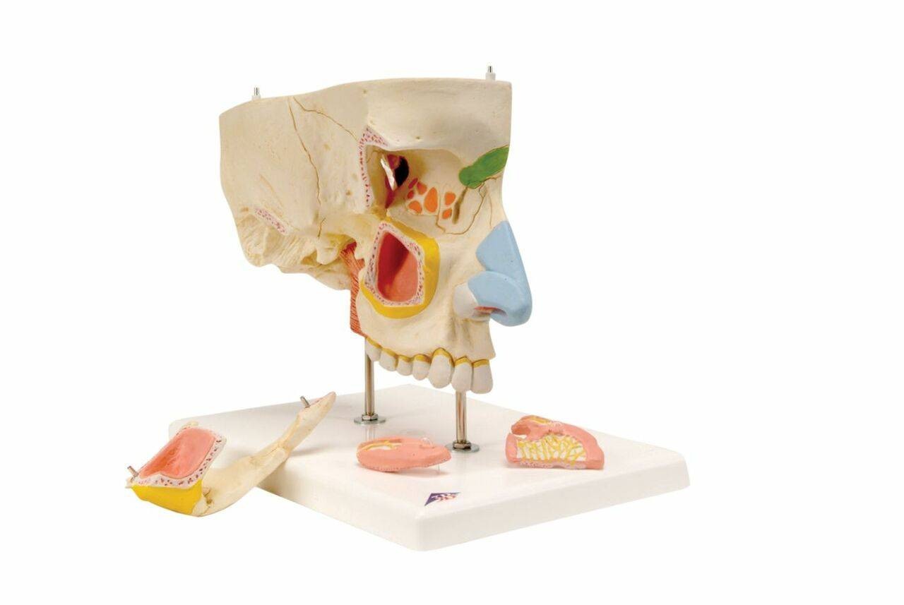
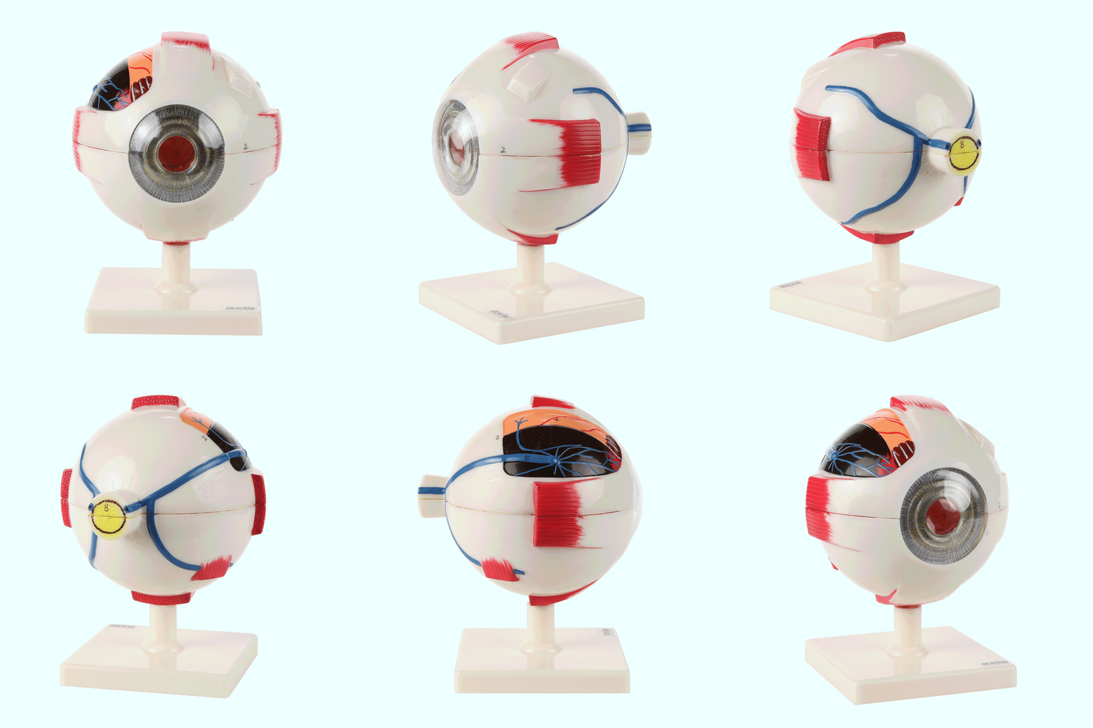
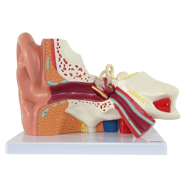
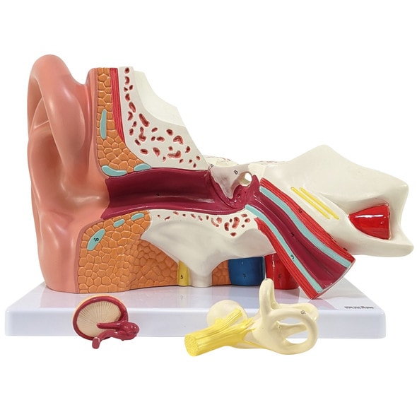
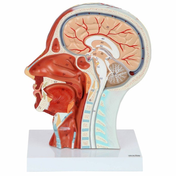
- Each model highlights essential internal structures and sensory receptors
- Perfect for high school science labs, undergraduate courses, and health education
- Durable construction built for repeated use in active learning environments
- Encourages visual, hands-on engagement with anatomical systems
Features Designed for Clarity and Comprehension
- Color-coded and labeled structures for simplified identification
- Removable or sectioned parts for internal view of organs
- Mounted on bases for easy handling and presentation
- Crafted with medical-grade PVC for durability
- Ideal for group learning or one-on-one instruction
Technical Specifications of the Product
- Product dimensions: Varies by model; each average 4–6 inches in height
- Product weight: Approx. 5 lbs total
- Product accessory specifics: All models include integrated display bases
- Included with purchase:
- 1 x Eye Model
- 1 x Ear Model
- 1 x Skin Cross-Section Model
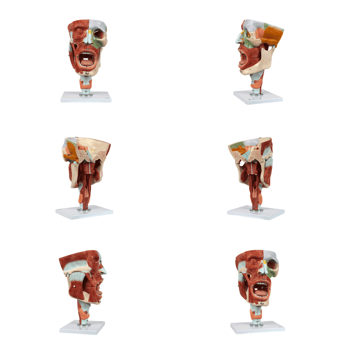
- 1 x Partial Jaw Model
- 1 x Product Manual with labeled diagrams

