Description
Explore the Depths of Human Anatomy—In Stunning 3D Detail
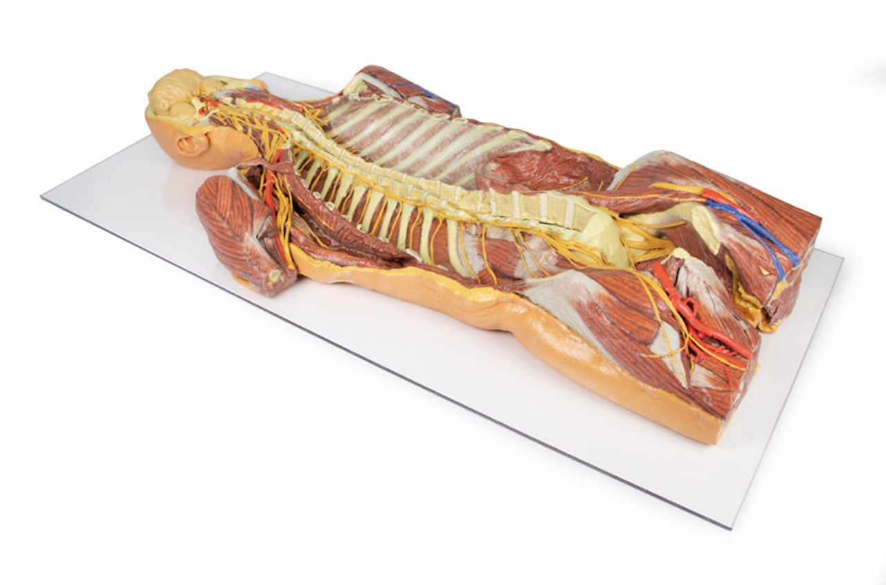
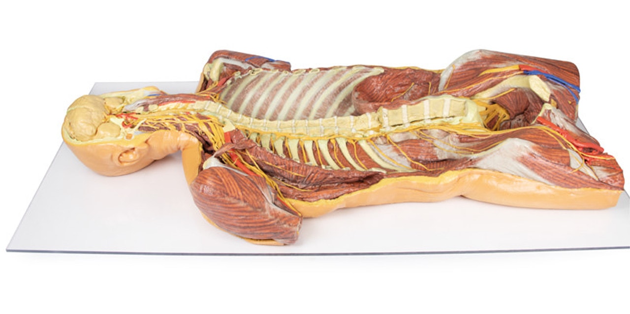
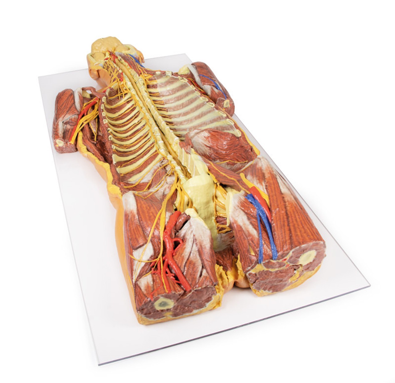
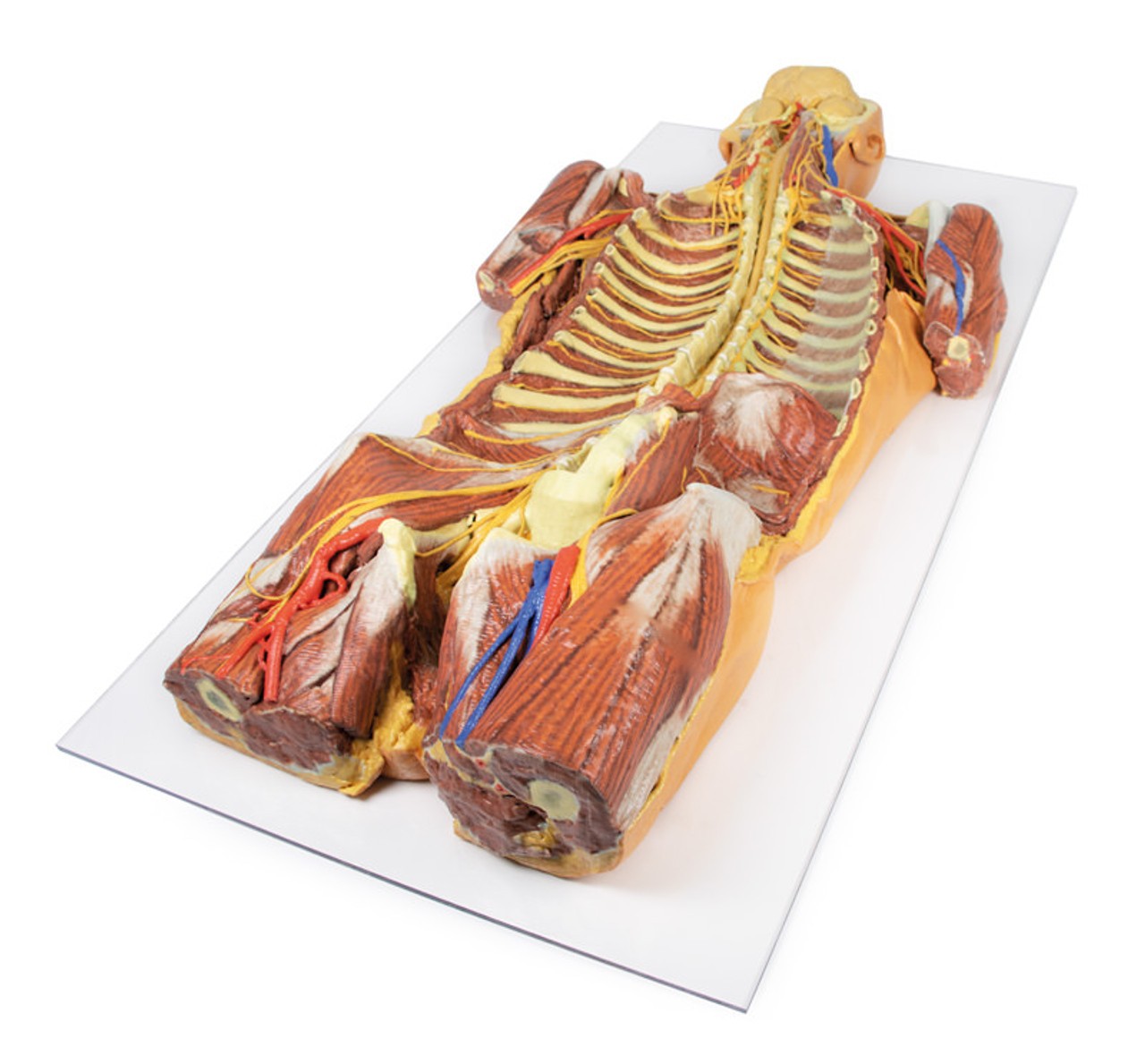
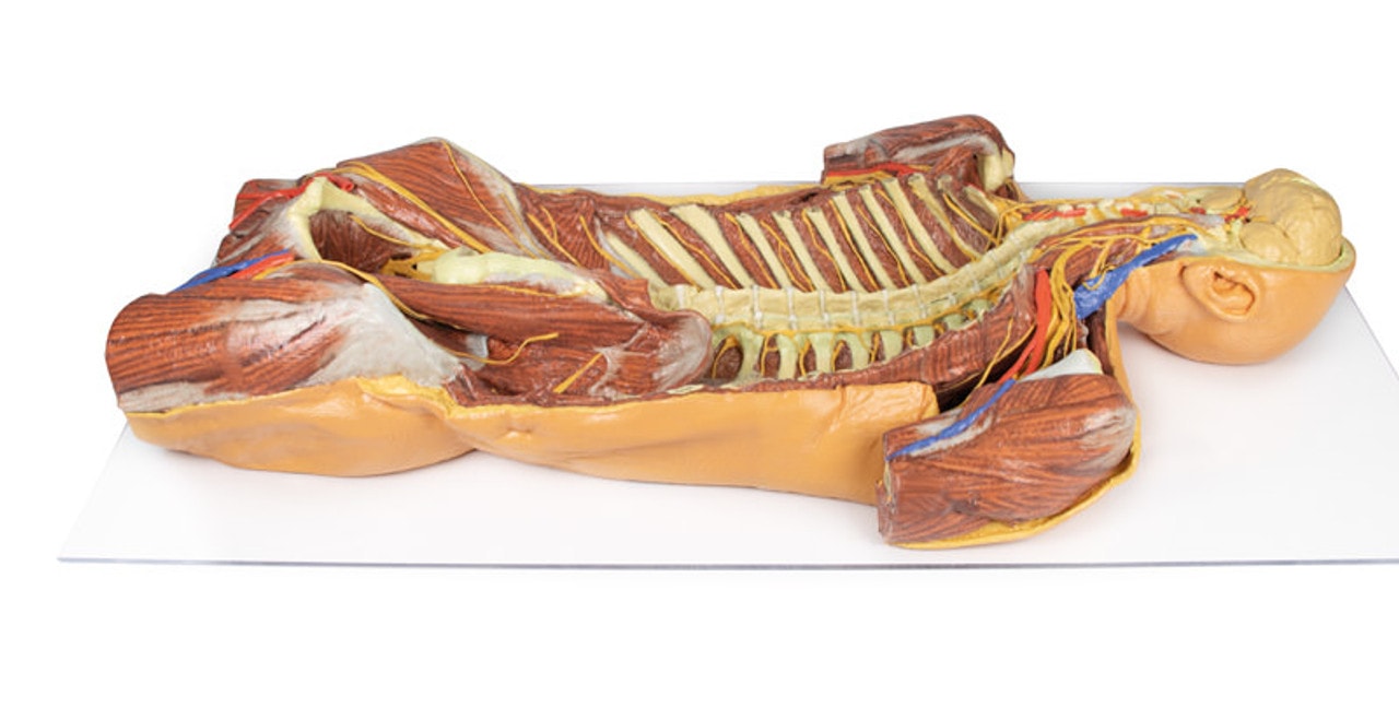
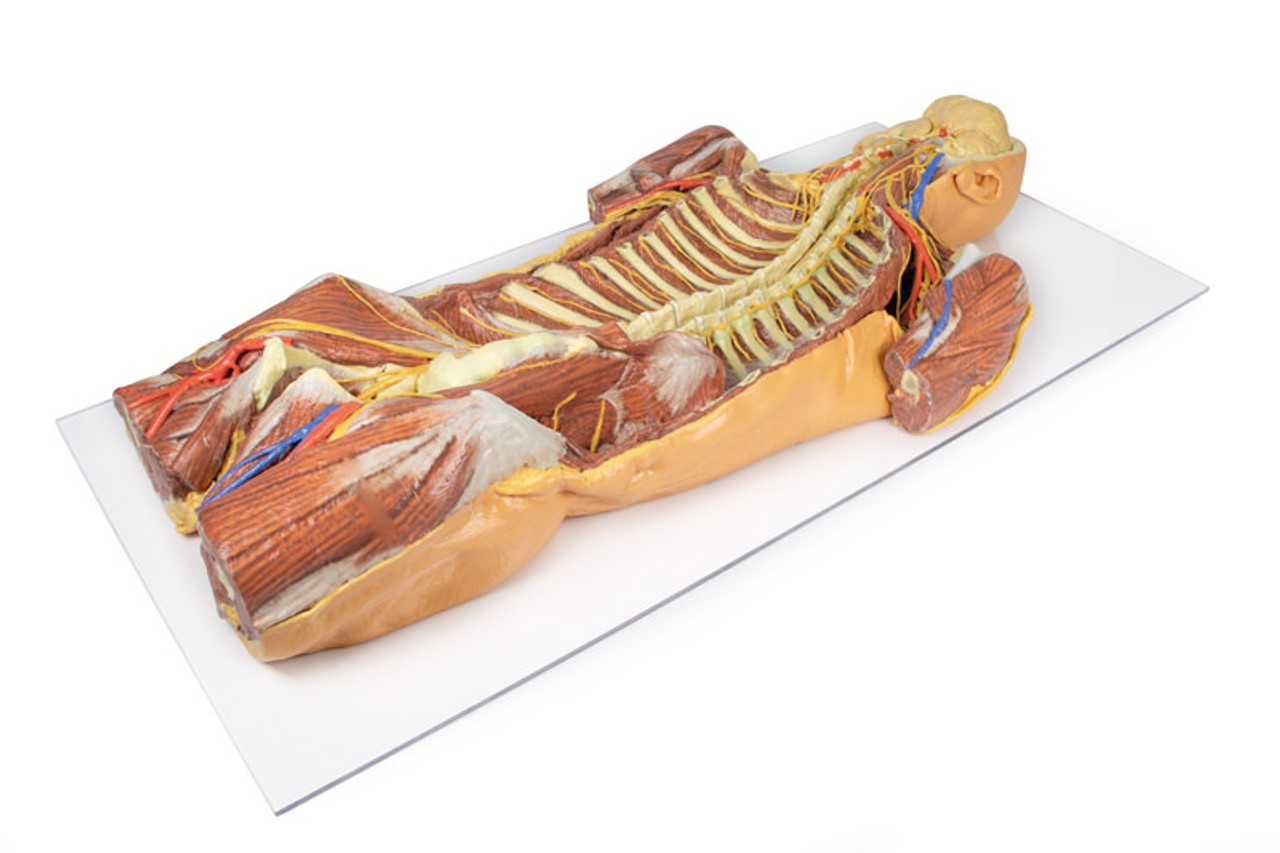
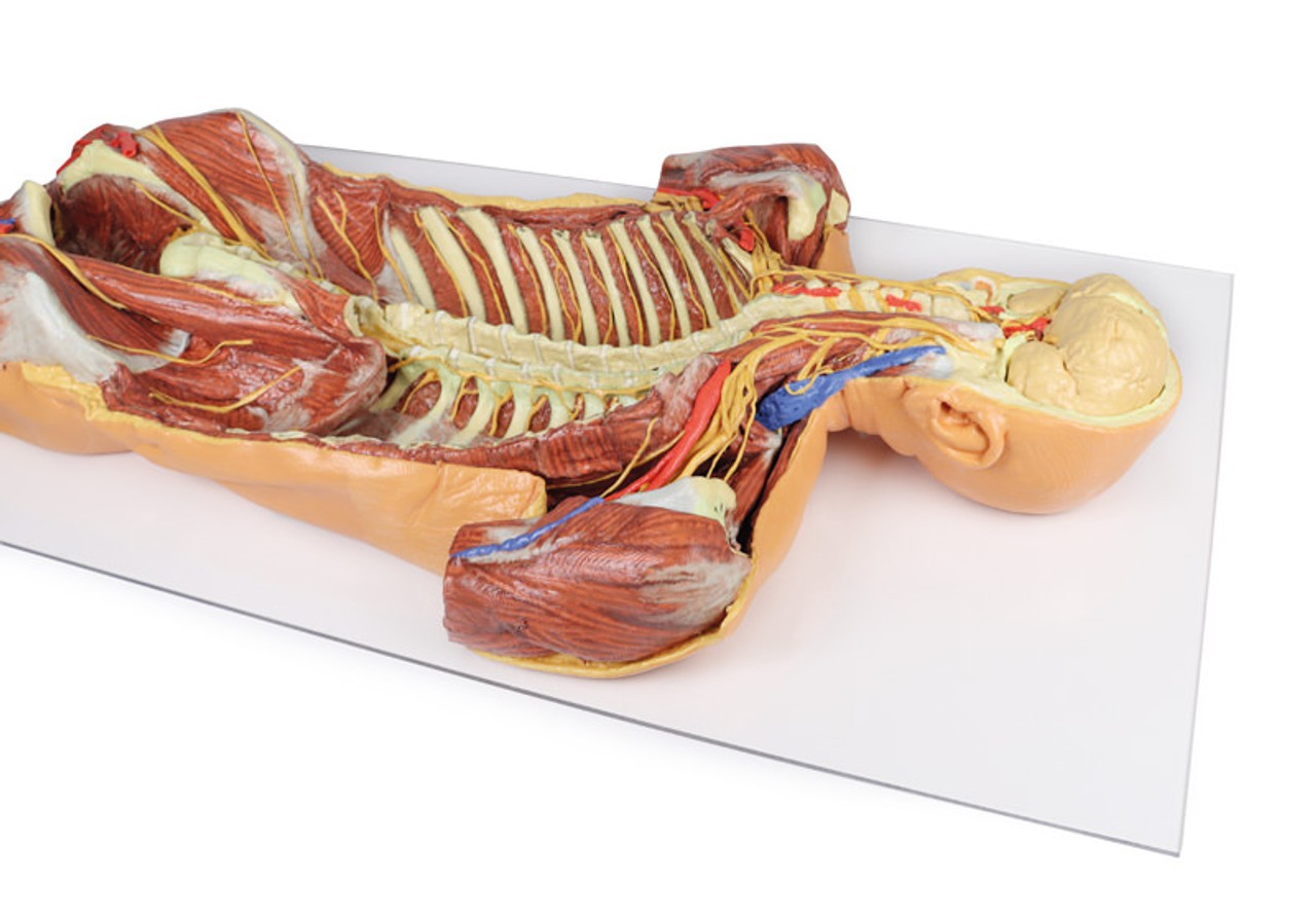
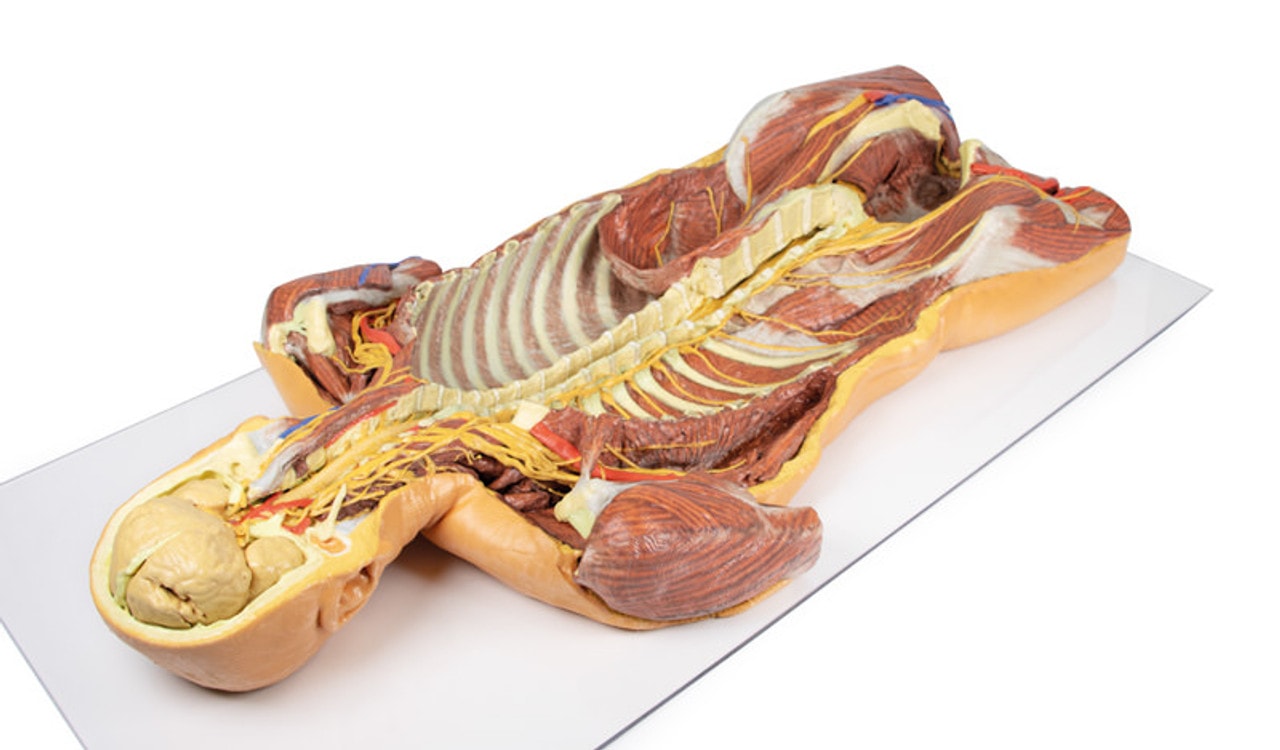
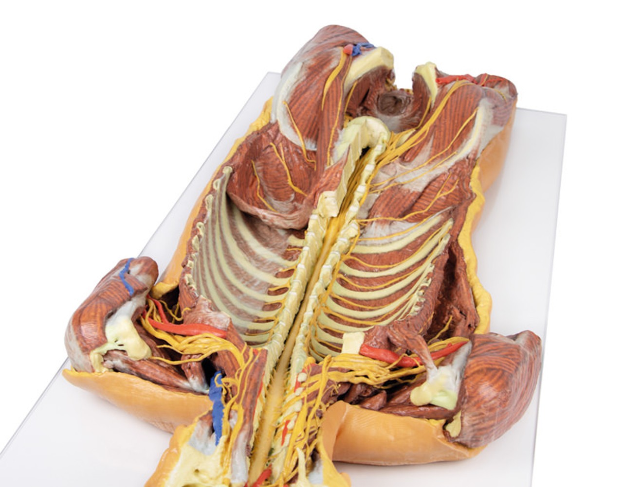
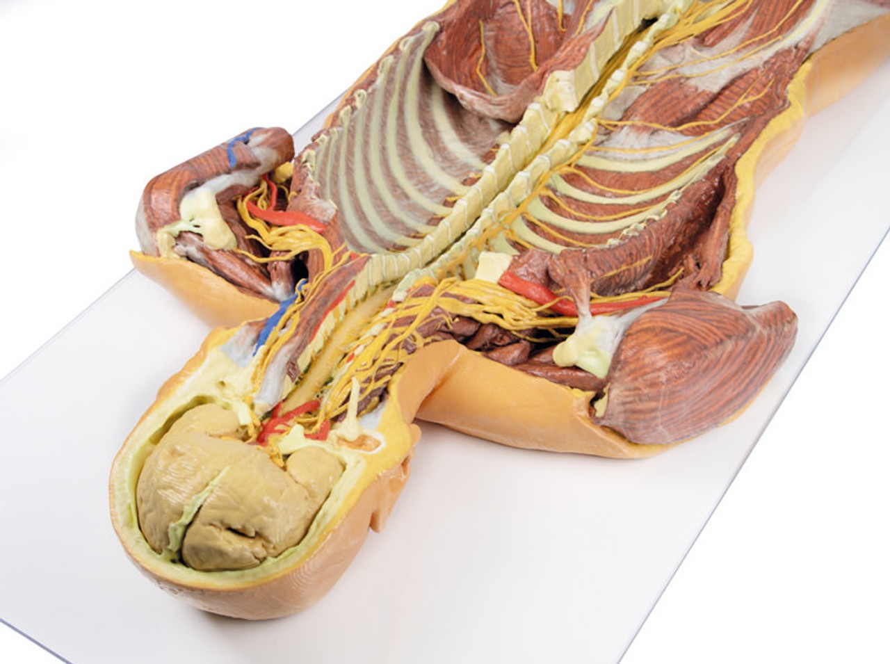
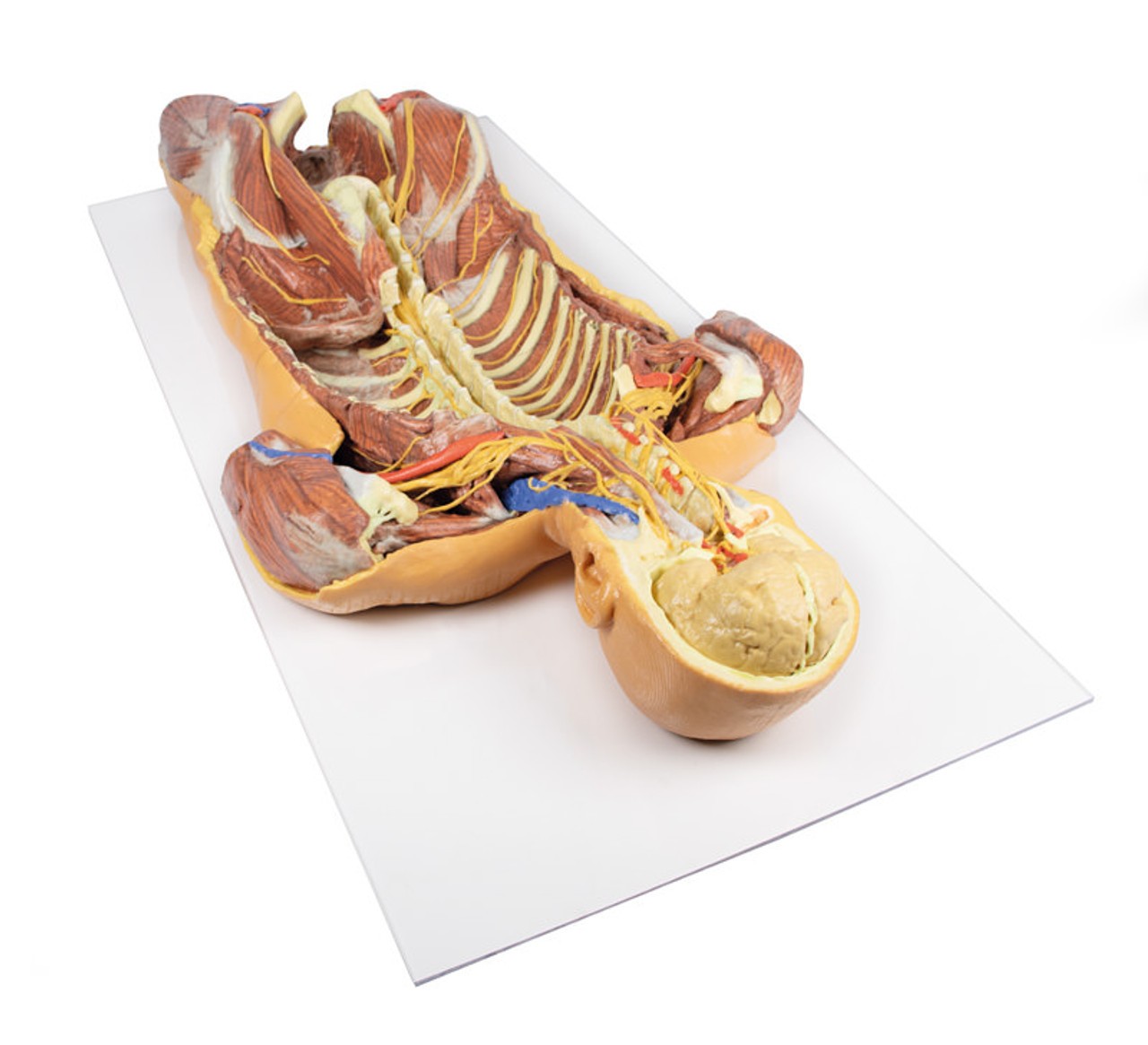
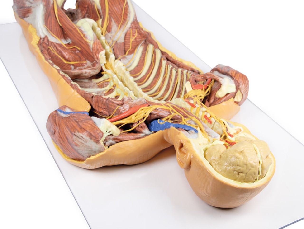
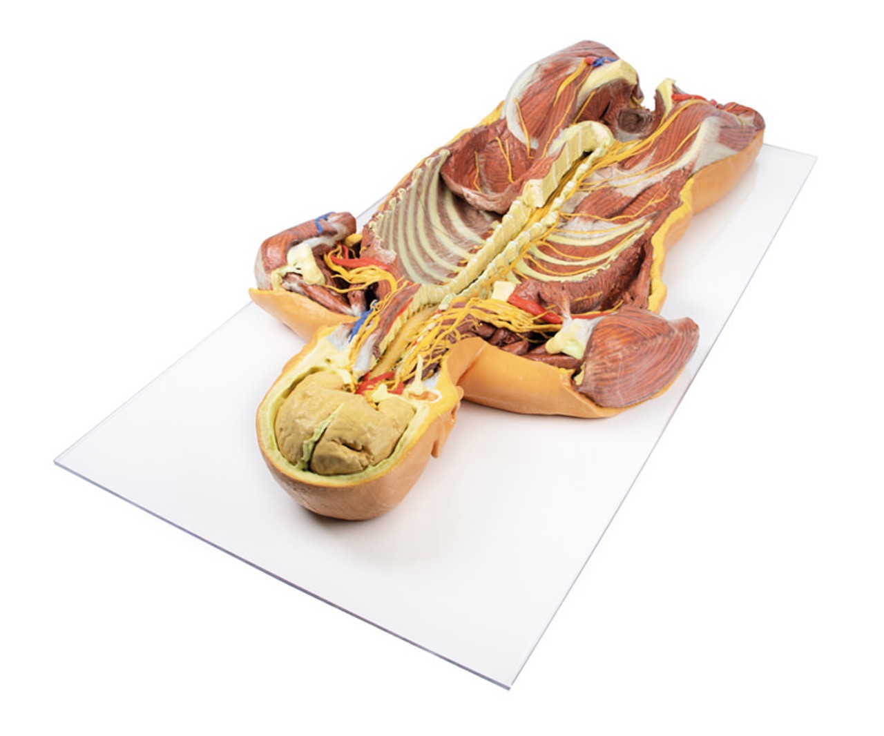
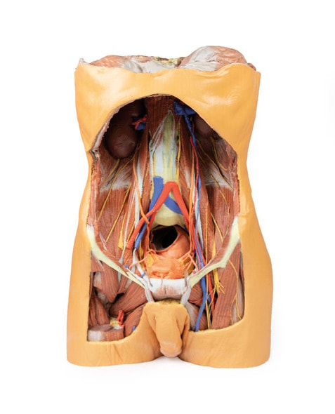
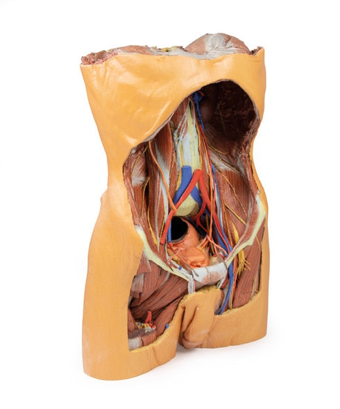
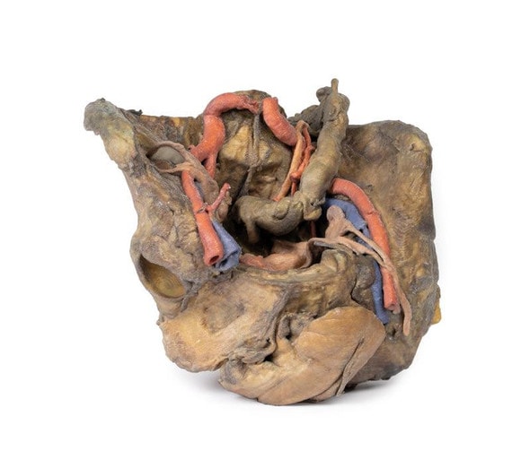
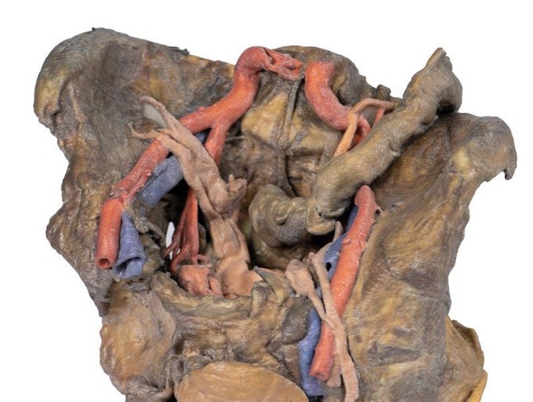
The 3D Printed Ventral Deep Dissection - Posterior Body Wall is a museum-quality model that captures a full-color, high-resolution anatomical dissection of the lower thoracic and abdominal cavities. Perfectly preserved and printed from real cadaveric data, this model offers an unprecedented view into the intricacies of the human body's core structures. Whether you're a medical student, educator, or clinical professional, this model will elevate your anatomical understanding. Don't miss the opportunity to bring this striking educational piece into your learning space today.
Built for Advanced Learning in Human Anatomy and Surgical Education
This model is designed for in-depth instruction of abdominal and posterior thoracic wall anatomy, showcasing internal organs, musculature, and vascular structures. It's particularly effective for teaching medical students, surgical residents, and anatomy scholars who require an accurate and tangible view of complex human systems. Use it to identify fascial layers, blood vessels, lymph nodes, and musculoskeletal relationships in precise anatomical context. From textbook theory to clinical practice, this model bridges the gap with stunning clarity.
A Glimpse Inside the Human Body—No Scalpel Required
- Visually striking full-color 3D print of real human dissection, derived from CT and photogrammetry scanning.
- Displays a partial vertebral column, exposed diaphragm, kidneys, psoas major, iliacus, and major vascular branches.
- Highlights cross-sectional structures like the liver, inferior vena cava, and abdominal aorta in spatial context.
- Useful for surgical planning, anatomical instruction, and procedural simulation environments.
- Ideal for medical schools, anatomy labs, simulation centers, and museum-quality display collections.
High-Impact Features for Realistic and Lasting Use
- Created from a real cadaver using high-resolution digital capture techniques
- Printed in full color using advanced 3D printing technology
- Durable polymer for long-term classroom or display use
- Detailed representation of soft tissue, musculature, and vasculature
- Comes with labeled anatomical guide for easy study and reference
Technical Specifications of the Product
- Product dimensions: 12 in x 24 x 47 in.
- Product weight: 5.5 lbs.
- Included with purchase:
- 1 x 3D Printed Ventral Deep Dissection - Posterior Body Wall

