Description
Developed from real patient case study specimens, the 3D printed anatomy model pathology series introduces an unmatched level of realism in human anatomy models. Each 3D printed anatomy model is a high-fidelity replica of a human cadaveric specimen, focusing on the key morbidity presentations that led to the deceasement of the patient. With advances in 3D printing materials and techniques, these stories can come to life in an ethical, consistently reproduceable, and easy to handle format. Ideal for the most advanced anatomical and pathological study, and backed by authentic case study details, students, instructors, and experts alike will discover a new level of anatomical study with the 3D printed anatomy model pathology series.
Clinical History
A 64-year old male presents with a 5-month history of generalized malaise, weight loss and dull right flank pain. On examination, there is a palpable right sided abdominal mass. He is noted to be hypertensive. Urinalysis reveals microscopic hematuria. The patient underwent right nephrectomy.
Pathology
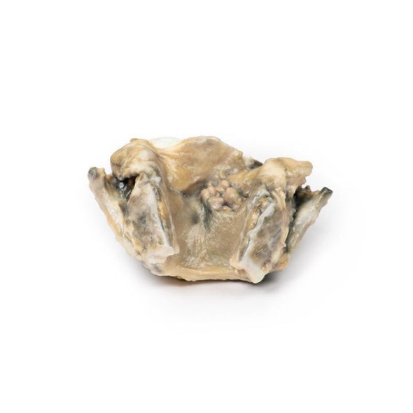
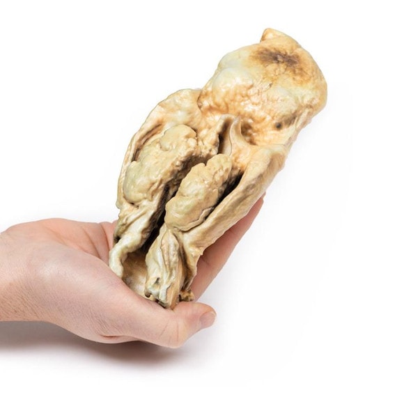
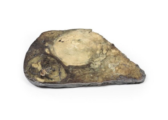
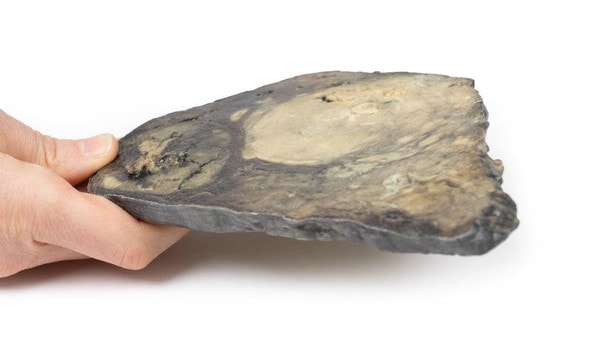
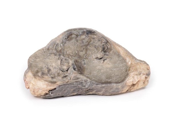
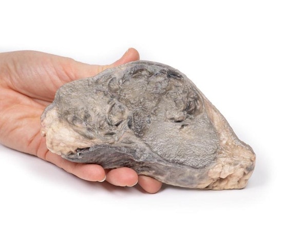
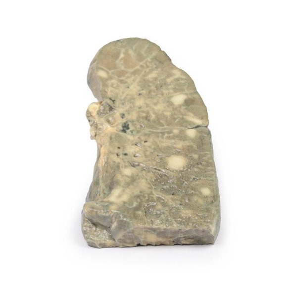
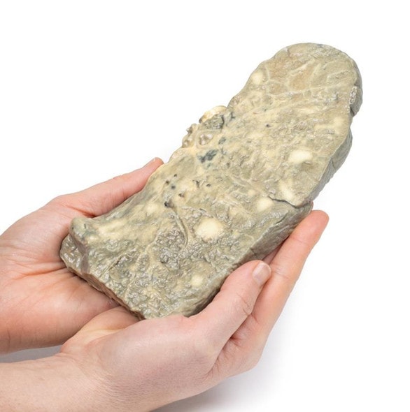
The specimen is a kidney, which has been incompletely dissected in the coronal plane, and mounted to display the cut surface. The lower pole of the kidney has been replaced by a rounded ill-defined irregular mass 5cm in diameter, which has compressed and distorted the overlying renal parenchyma. The cut surface of the tumor has a variegated appearance caused by areas of hemorrhage and necrosis. Several small pale-yellow tumor nodules are present in the cortex and medulla above and separate from the lower pole tumor. These are intrarenal metastases. The renal pelvis appears slightly dilated with some blunting of the renal papillae, suggesting a degree of hydronephrosis. The capsular surface is finely nodular with a few coarse scars and contains several small simple cysts (see rear of specimen). Histologically, the tumor was diagnosed as a renal cell carcinoma.
Further Information
Renal cell carcinoma (RCC) comprise 85% of the primary renal malignancies. They originate within the renal cortex. The risk of developing RCC is doubled in males. It most commonly occurs in the 6th decade of life. Other risk factors for RCC include smoking, obesity, hypertension, unopposed estrogen therapy, as well as exposure to asbestos, petroleum and heavy metals. Most RCC are sporadic but around 5% are due to autosomal dominant familial cancers, such as Von Hippel Lindau syndrome, hereditary leiomyomatosis and Birt-Hogg-Dub syndrome.
There are several major primary renal tumor types according to genetic and histological tumor characteristics: clear cell carcinoma (70-80%), papillary carcinoma (10-15%), chromophobe carcinoma (5-10%), oncytic carcinoma (3-7%) and collecting (Bellini) Duct carcinoma (<1%).
Clear cell carcinoma typically have a deletion of chromosome 3p and arise from the proximal tubule. They may be solid or less commonly cystic. They occur in association with Von Hippel Lindau as well as sporadically. Papillary carcinomas arise from the proximal tubule. They are associated with trisomies 7 and 17; loss of Y in male patients; and MET kinase domain mutations. They are frequently multifocal in origin. Chromophobe carcinoma originate from intercalated cells of the collecting ducts. They are associated with multiple chromosome losses and hypodiploidy. They have a low risk of disease progression.
Renal oncocytic carcinomas are typically comprised of well-differentiated cells with prominently eosinophilic granular cytoplasm; they are associated with a good prognosis. In contrast, collecting (Bellini) duct carcinoma of the kidney is a highly aggressive tumor with an extremely poor prognosis as it does not respond well to chemotherapy drugs used for renal cell carcinoma, and progresses and spreads more quickly. It is a variety of renal cell carcinoma (RCC) arising from the distal segment of the collecting ducts of Bellini in the renal medulla.
The typical clinical features of RCC are costovertebral pain, palpable mass and hematuria. RCC is the great mimic in medicine producing many manifestations including: polycythemia, hypercalcemia, hypertension, pyrexia, Cushing's syndrome, eosinophilia and amyloidosis. RCC tend to metastasize before producing may local symptoms. The most common sites of distal spread are the lungs (50%) and bones (33%) followed by lymph nodes, adrenal glands and brain. RCC has a tendency to invade the renal vein and extend up it as a tumor thrombus, growing as a solid column extending up to the inferior vena cava.
Ultrasound and CT are the most common investigations used to assess renal lesions and diagnose RCC. A tissue biopsy may be required in some patients. An increasing number of patients are being diagnosed with RCC because of incidental kidney lesions being detected on abdominal CT requested for other medical reasons.
The average 5-year survival rate for RCC is 70%. Treatment depends on the stage of the tumor. Radical nephrectomy is the usual surgical option. Medical treatment includes chemotherapeutic drugs as well as Vascular endothelial growth factor (VEGF) inhibitors and tyrosine kinases inhibitors in patients with metastatic disease.
Advantages of 3D Printed Anatomical Models
- 3D printed anatomical models are the most anatomically accurate examples of human anatomy because they are based on real human specimens.
- Avoid the ethical complications and complex handling, storage, and documentation requirements with 3D printed models when compared to human cadaveric specimens.
- 3D printed anatomy models are far less expensive than real human cadaveric specimens.
- Reproducibility and consistency allow for standardization of education and faster availability of models when you need them.
- Customization options are available for specific applications or educational needs. Enlargement, highlighting of specific anatomical structures, cutaway views, and more are just some of the customizations available.
Disadvantages of Human Cadavers
- Access to cadavers can be problematic and ethical complications are hard to avoid. Many countries cannot access cadavers for cultural and religious reasons.
- Human cadavers are costly to procure and require expensive storage facilities and dedicated staff to maintain them. Maintenance of the facility alone is costly.
- The cost to develop a cadaver lab or plastination technique is extremely high. Those funds could purchase hundreds of easy to handle, realistic 3D printed anatomical replicas.
- Wet specimens cannot be used in uncertified labs. Certification is expensive and time-consuming.
- Exposure to preservation fluids and chemicals is known to cause long-term health problems for lab workers and students. 3D printed anatomical replicas are safe to handle without any special equipment.
- Lack of reuse and reproducibility. If a dissection mistake is made, a new specimen has to be used and students have to start all over again.
Disadvantages of Plastinated Specimens
- Like real human cadaveric specimens, plastinated models are extremely expensive.
- Plastinated specimens still require real human samples and pose the same ethical issues as real human cadavers.
- The plastination process is extensive and takes months or longer to complete. 3D printed human anatomical models are available in a fraction of the time.
- Plastinated models, like human cadavers, are one of a kind and can only showcase one presentation of human anatomy.
Advanced 3D Printing Techniques for Superior Results
- Vibrant color offering with 10 million colors
- UV-curable inkjet printing
- High quality 3D printing that can create products that are delicate, extremely precise, and incredibly realistic
- To improve durability of fragile, thin, and delicate arteries, veins or vessels, a clear support material is printed in key areas. This makes the models robust so they can be handled by students easily.


