Description
Median Section of a Dog's Head: A Detailed Description for Veterinary Education
Introduction
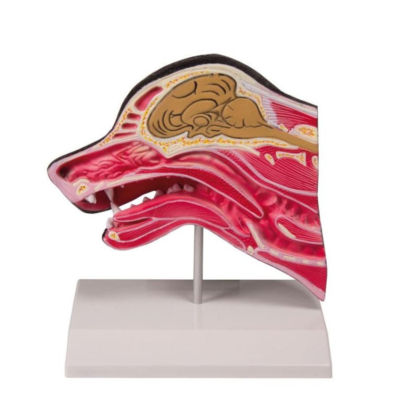
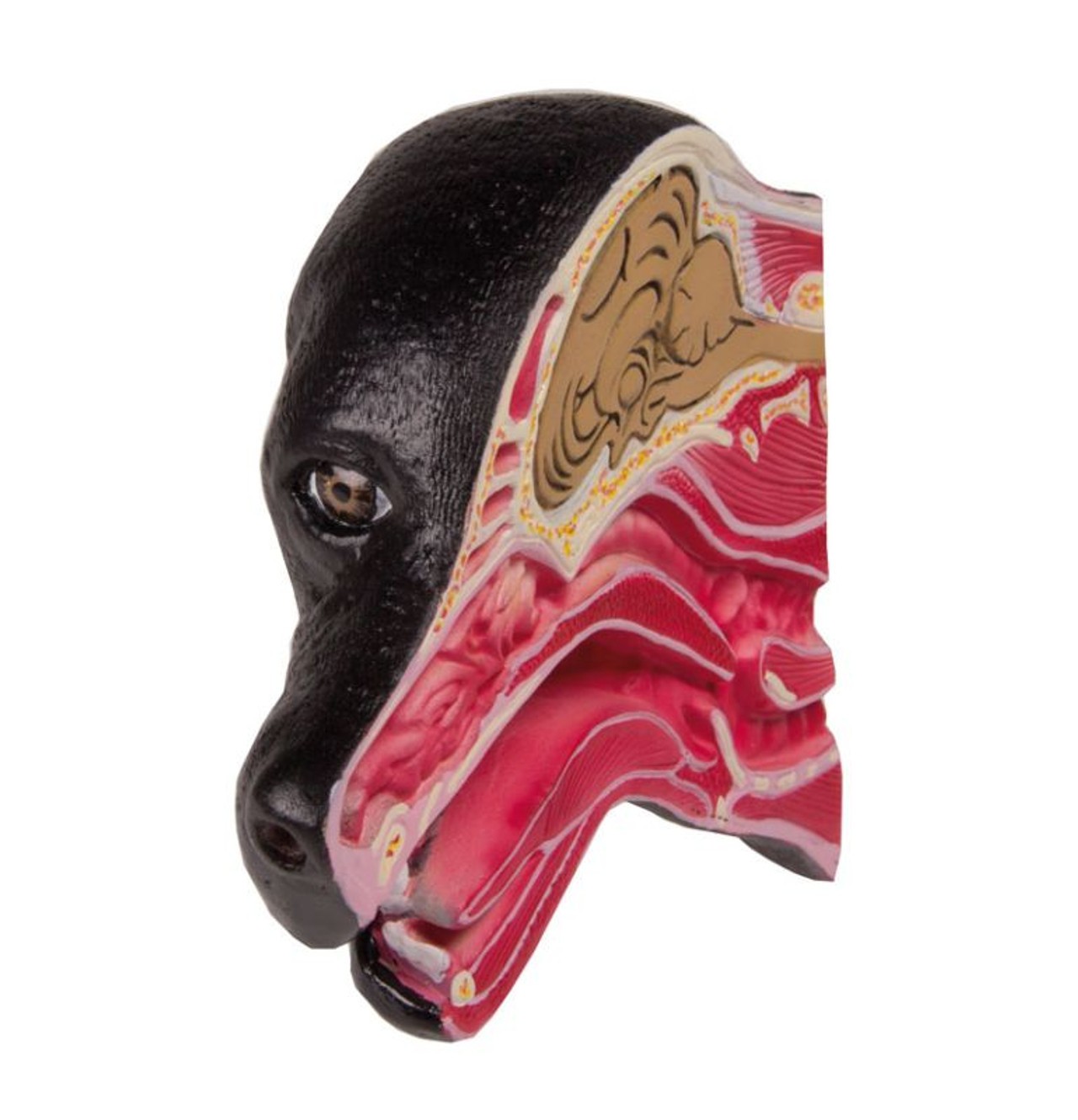
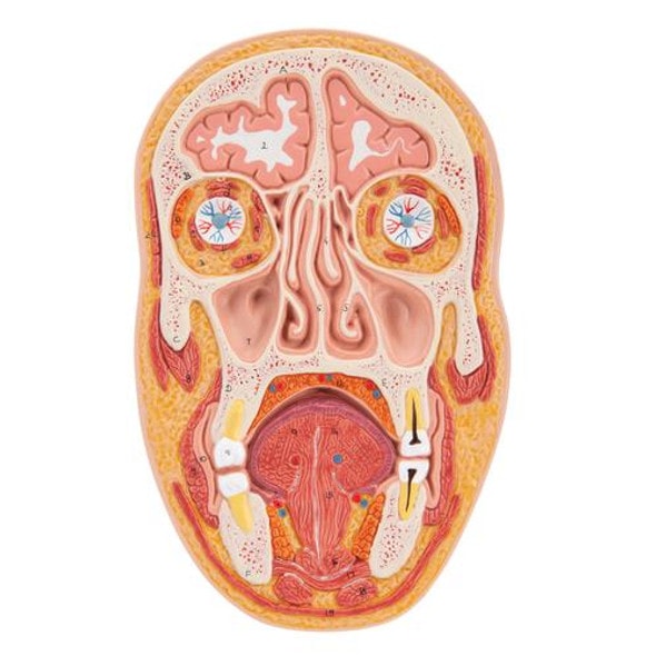
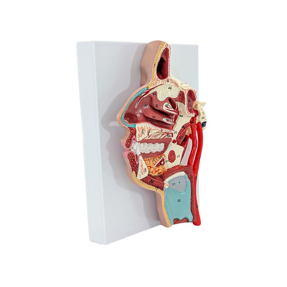
The median section of a dog's head provides crucial insights into the internal structures and their spatial relationships. This anatomical view is essential for veterinary students, technicians, and professionals for diagnostic, surgical, and clinical procedures. The median plane divides the head into left and right halves, offering a comprehensive view of the internal structures of the mouth, tongue, throat and skull.
1. External Features
- Nasal Cavity: The nasal cavity is situated between the nasal bones and is divided into two nostrils by the nasal septum. The median section reveals the nasal turbinates (conchae), which are important for filtering, warming, and humidifying inspired air.
- Oral Cavity: The oral cavity includes the maxilla and mandible. The median section exposes the palatine bones forming the hard palate and the soft palate. The section also shows the position of the teeth in the upper and lower jaws, including incisors, canines, premolars, and molars.
2. Skeletal Structures
- Skull: The median section of the skull includes the frontal bone, nasal bones, and the sphenoid bone. The section highlights the orbital cavities and the positioning of the eye sockets.
- Mandible and Maxilla: The mandible (lower jaw) and maxilla (upper jaw) are visible in this section, showing the alignment of the teeth and the articulation of the jaw.
3. Muscles
- Masticatory Muscles: The median section allows visualization of the primary muscles involved in mastication, such as the masseter, temporalis, and pterygoid muscles. Understanding their attachment sites and functions is crucial for evaluating bite force and jaw function.
- Facial Muscles: The section also reveals facial muscles such as the orbicularis oris and buccinator, which are involved in facial expression and the movement of the lips and cheeks.
4. Neural Structures
- Brain: The median section includes parts of the brain such as the cerebrum, cerebellum, and brainstem.
- Sinuses and Eustachian Tube: The section shows the frontal and maxillary sinuses and the Eustachian tube, which connects the middle ear to the nasopharynx.
5. Vascular Structures
- Major Arteries and Veins: The median section reveals the position of the major arteries such as the common carotid arteries and their branches. It also shows major veins, including the internal jugular veins, which are important for understanding blood flow and diagnosing vascular conditions.
6. Other Structures
- Pharynx and Larynx: The median section displays the nasopharynx and the oropharynx, as well as the larynx. These structures are vital for understanding the airway and vocalization mechanisms.
- Esophagus: The esophagus can be seen posterior to the trachea, providing insight into the digestive tract's alignment and function.
Conclusion
The median section of a dog's head offers a comprehensive view of the internal anatomy, essential for veterinary education and practice. Understanding this section aids in diagnosing medical conditions, planning surgical interventions, and performing detailed anatomical studies. For veterinary students and professionals, this detailed view is indispensable for developing a thorough knowledge of canine anatomy.

