Description
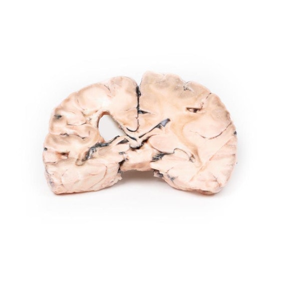
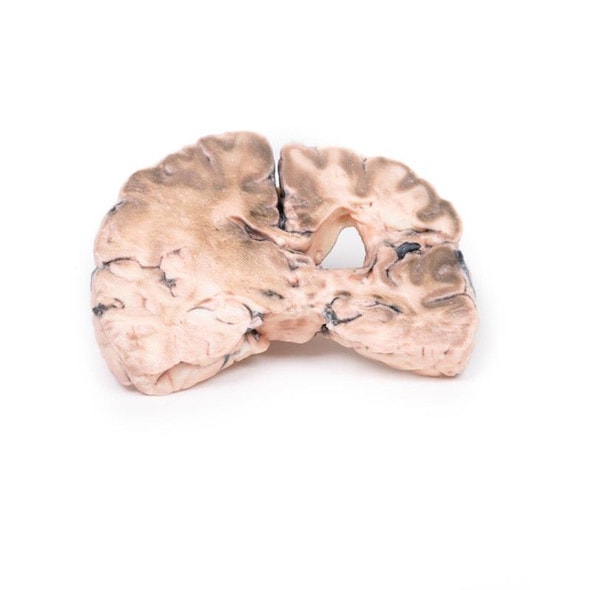
Developed from real patient case study specimens, the 3D printed anatomy model pathology series introduces an unmatched level of realism in human anatomy models. Each 3D printed anatomy model is a high-fidelity replica of a human cadaveric specimen, focusing on the key morbidity presentations that led to the deceasement of the patient. With advances in 3D printing materials and techniques, these stories can come to life in an ethical, consistently reproduceable, and easy to handle format. Ideal for the most advanced anatomical and pathological study, and backed by authentic case study details, students, instructors, and experts alike will discover a new level of anatomical study with the 3D printed anatomy model pathology series.
Clinical History
A 54 year old male patient presents with flank pain. He is an active intravenous drug user. Further questioning reveals a history of intermittent hematuria, fevers, malaise and vomiting. On examination he is hypertensive and pyrexic. Inspection of his limbs reveals Janeway lesions on his extremities and track marks from recent IV drug use. A systolic murmur is found on auscultation of his chest. Blood tests reveal elevated inflammatory markers, impaired renal function, elevated LDH and multiple bacteremia blood cultures. Echocardiogram shows a large mobile tricuspid vegetation. He was commenced on treatment for infective endocarditis but later died from a sudden cardiac arrest.
Pathology
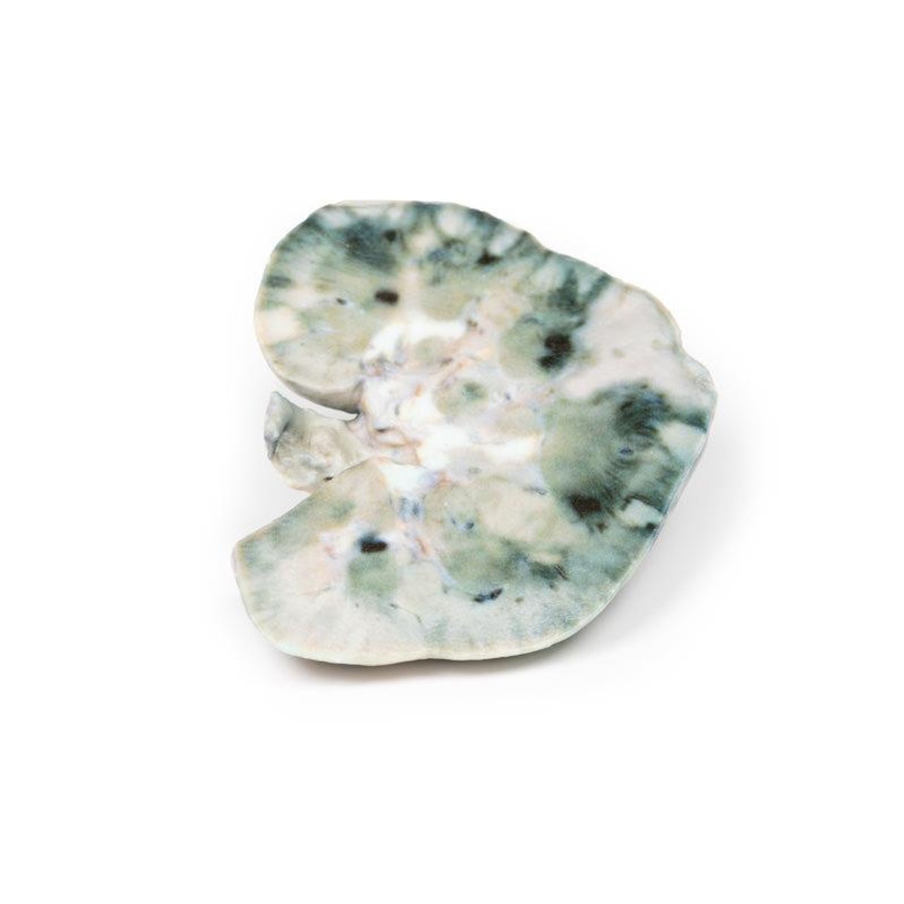
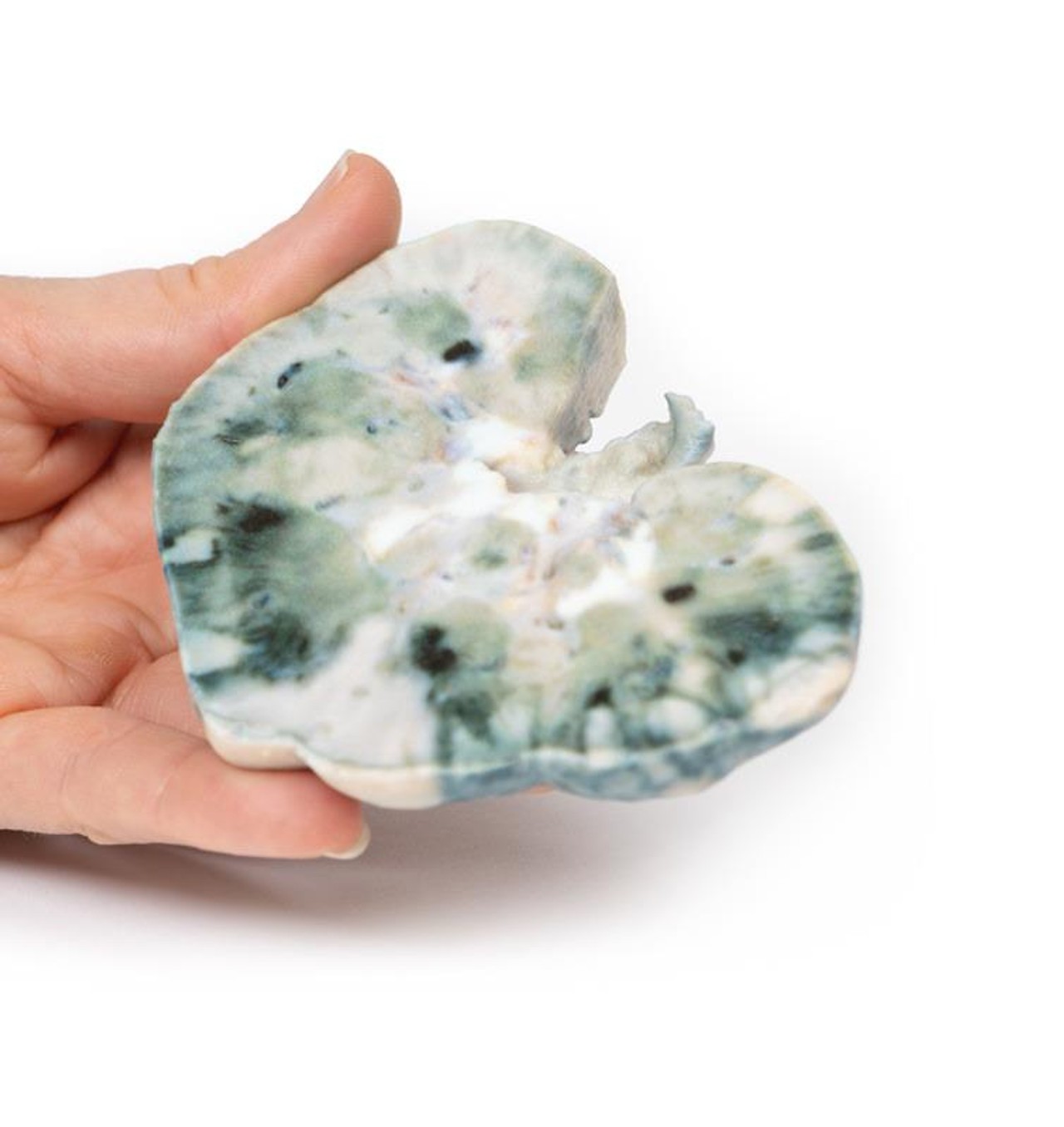
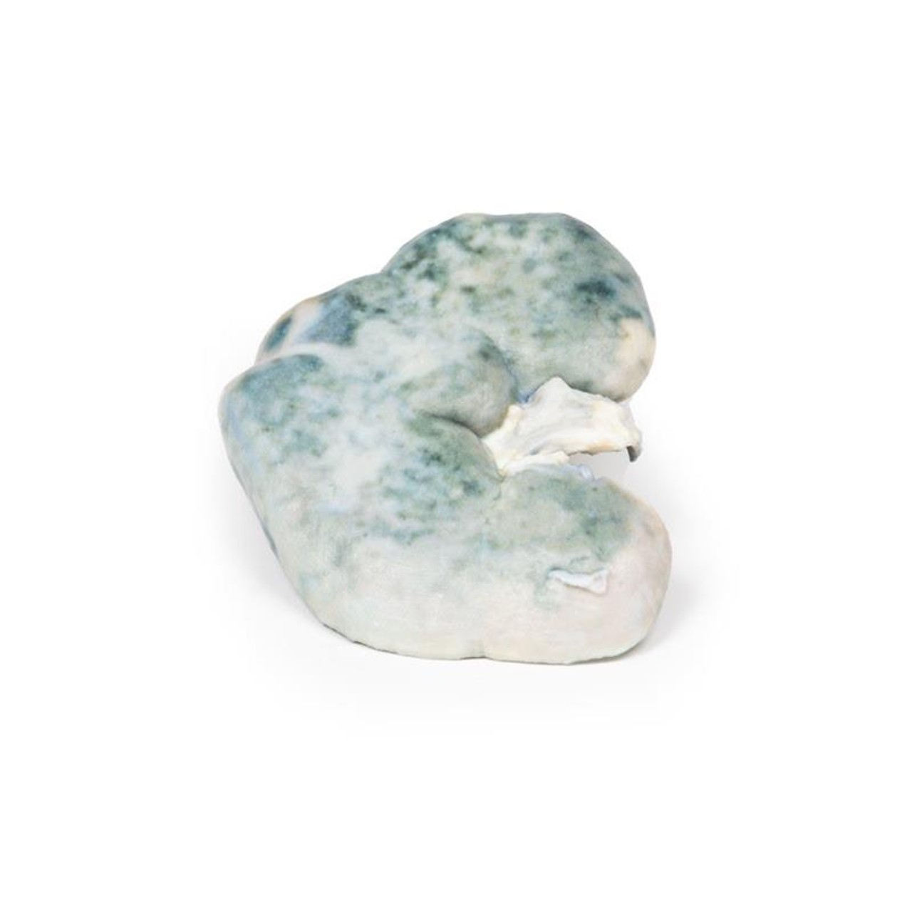
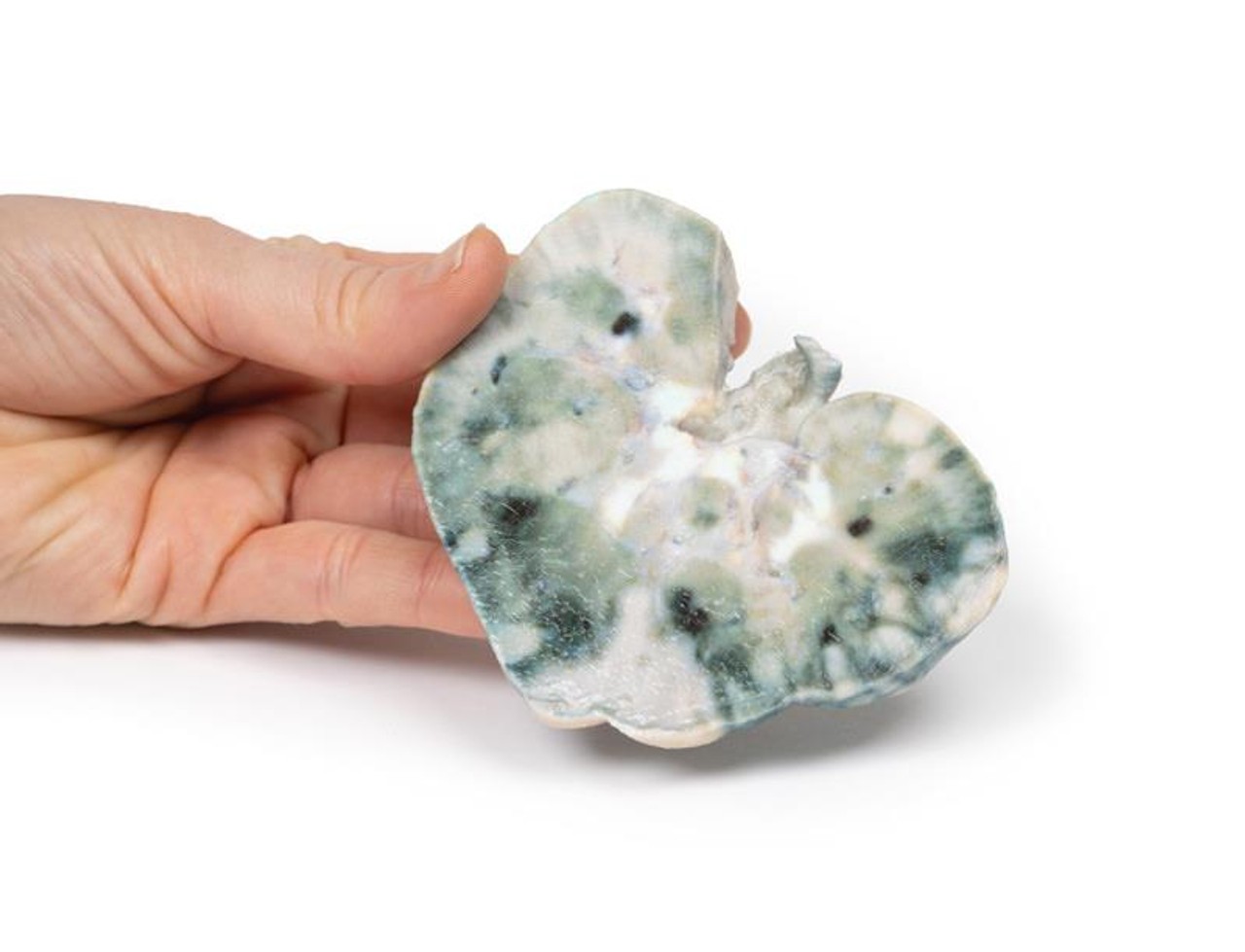
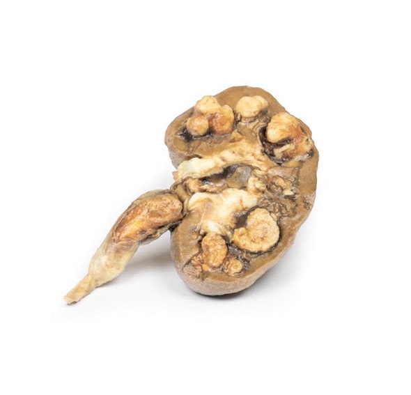
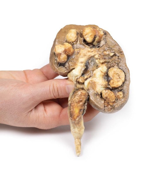
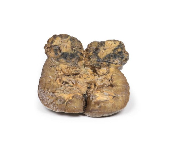
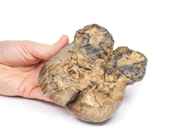
The specimen is the patient's kidney from post mortem examination. The kidney has been bisected with a cut half surface on display. There are multiple well demarcated wedge shaped pale yellow-white areas evident within the cortex. The base of these pyramids lies against the cortical surface and extend along the renal columns with the apex pointing toward the medulla. The largest is evident lateral upper pole of the kidney. Theses pale areas are infarcted renal tissue. There are dark irregular shaped areas which represent areas of hemorrhage.
Further Information
Renal infarction results from an interruption in the blood flow to the kidney. The kidneys receive almost a quarter of the cardiac output but have limited collateral circulation. The cortex is the most susceptible area to infarction given blood supply is from proximal to distal. The main causes of interruption of this circulation are cardioembolic disease, renal artery damage, hypercoagulable states or idiopathic.
Cardioembolic causes are the most common. These include post-myocardial infarction mural thrombi, septic emboli from infective endocarditis and emboli from mechanical valves. Idiopathic renal infarction is the second most common cause. Damage to the renal artery is the third most frequent cause and includes renal artery dissection, acute vasculitis of polyarteritis nodosa, trauma or post endovascular intervention. Hypercoagulable states are the rarest cause of renal infarcts such as hereditary thrombophilia and antiphospholipid syndrome. Infarction is bilateral in ~15% of cases.
Presentation of renal infarction depends on the underlying etiology. It can be clinically silent. Common manifestations include costovertebral angle pain, hematuria, hypertension due to increased renin release, nausea, vomiting and sometimes fever.
Laboratory test used to aid diagnosis include urinalysis for hematuria and serum creatinine levels which may be elevated, especially in bilateral disease. CT abdomen with contrast is the first choice radiological investigation. A wedge-shaped perfusion defect is the classic finding. Treatment varies depending on the cause of the infarction but generally involves supportive therapy and treatment of the underlying pathology.
Advantages of 3D Printed Anatomical Models
- 3D printed anatomical models are the most anatomically accurate examples of human anatomy because they are based on real human specimens.
- Avoid the ethical complications and complex handling, storage, and documentation requirements with 3D printed models when compared to human cadaveric specimens.
- 3D printed anatomy models are far less expensive than real human cadaveric specimens.
- Reproducibility and consistency allow for standardization of education and faster availability of models when you need them.
- Customization options are available for specific applications or educational needs. Enlargement, highlighting of specific anatomical structures, cutaway views, and more are just some of the customizations available.
Disadvantages of Human Cadavers
- Access to cadavers can be problematic and ethical complications are hard to avoid. Many countries cannot access cadavers for cultural and religious reasons.
- Human cadavers are costly to procure and require expensive storage facilities and dedicated staff to maintain them. Maintenance of the facility alone is costly.
- The cost to develop a cadaver lab or plastination technique is extremely high. Those funds could purchase hundreds of easy to handle, realistic 3D printed anatomical replicas.
- Wet specimens cannot be used in uncertified labs. Certification is expensive and time-consuming.
- Exposure to preservation fluids and chemicals is known to cause long-term health problems for lab workers and students. 3D printed anatomical replicas are safe to handle without any special equipment.
- Lack of reuse and reproducibility. If a dissection mistake is made, a new specimen has to be used and students have to start all over again.
Disadvantages of Plastinated Specimens
- Like real human cadaveric specimens, plastinated models are extremely expensive.
- Plastinated specimens still require real human samples and pose the same ethical issues as real human cadavers.
- The plastination process is extensive and takes months or longer to complete. 3D printed human anatomical models are available in a fraction of the time.
- Plastinated models, like human cadavers, are one of a kind and can only showcase one presentation of human anatomy.
Advanced 3D Printing Techniques for Superior Results
- Vibrant color offering with 10 million colors
- UV-curable inkjet printing
- High quality 3D printing that can create products that are delicate, extremely precise, and incredibly realistic
- To improve durability of fragile, thin, and delicate arteries, veins or vessels, a clear support material is printed in key areas. This makes the models robust so they can be handled by students easily.

