Description
Developed from real patient case study specimens, the 3D printed anatomy model pathology series introduces an unmatched level of realism in human anatomy models. Each 3D printed anatomy model is a high-fidelity replica of a human cadaveric specimen, focusing on the key morbidity presentations that led to the deceasement of the patient. With advances in 3D printing materials and techniques, these stories can come to life in an ethical, consistently reproduceable, and easy to handle format. Ideal for the most advanced anatomical and pathological study, and backed by authentic case study details, students, instructors, and experts alike will discover a new level of anatomical study with the 3D printed anatomy model pathology series.
Clinical History
A 10-year-old girl with a known congenital heart was admitted for surgical repair because of the recent onset of cyanosis and cardiac failure. On examination, she was breathless with a blood pressure of 105/60mm/Hg and a pulse rate of 140/min. There was a loud heart murmur in the fourth left intercostal space adjacent to the sternum. The jugular venous pressure was elevated, and there were bilateral pulmonary basal crepitations but no peripheral oedema. At operation, the defect was repaired; however, death followed a sudden post-operative deterioration of unknown cause.
Pathology
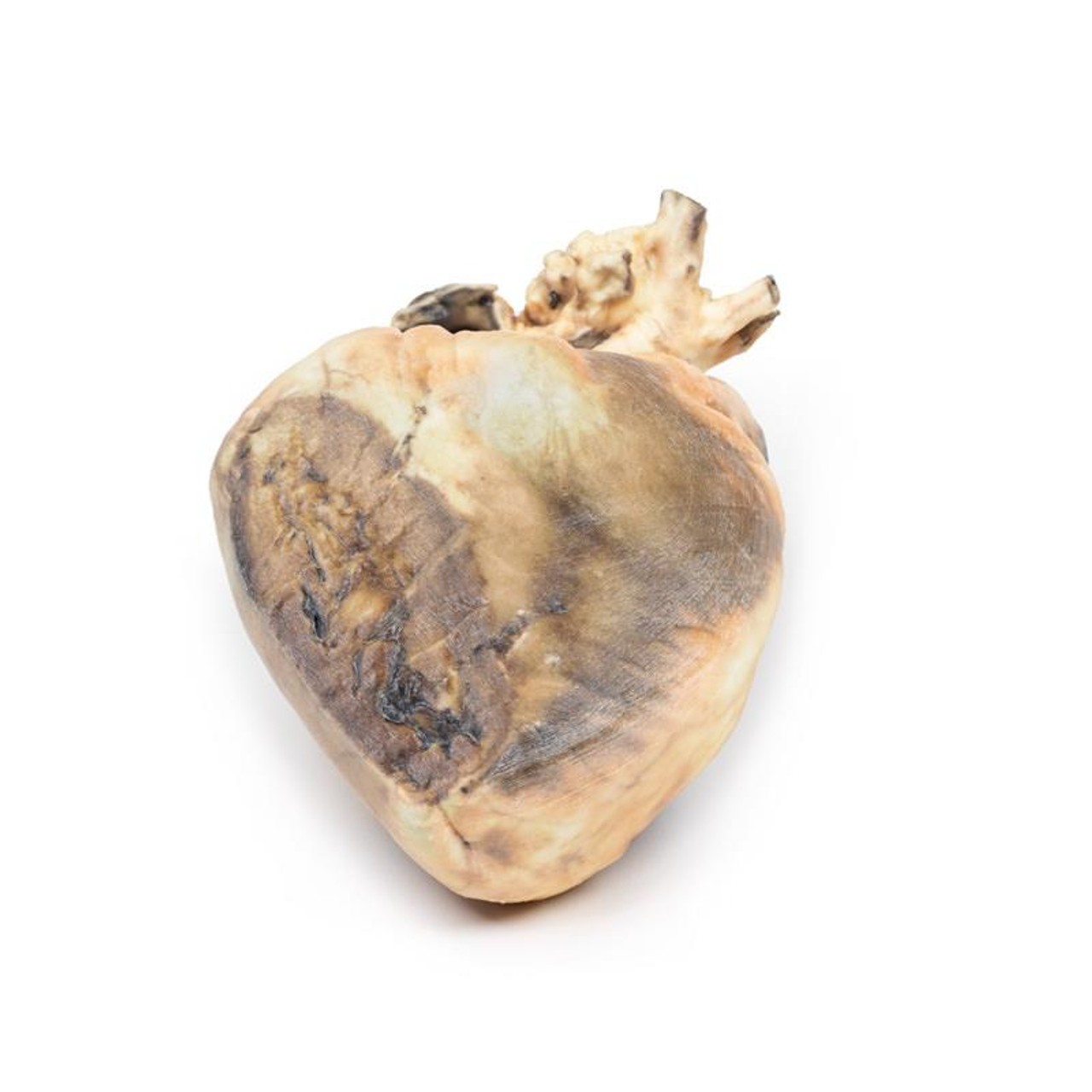
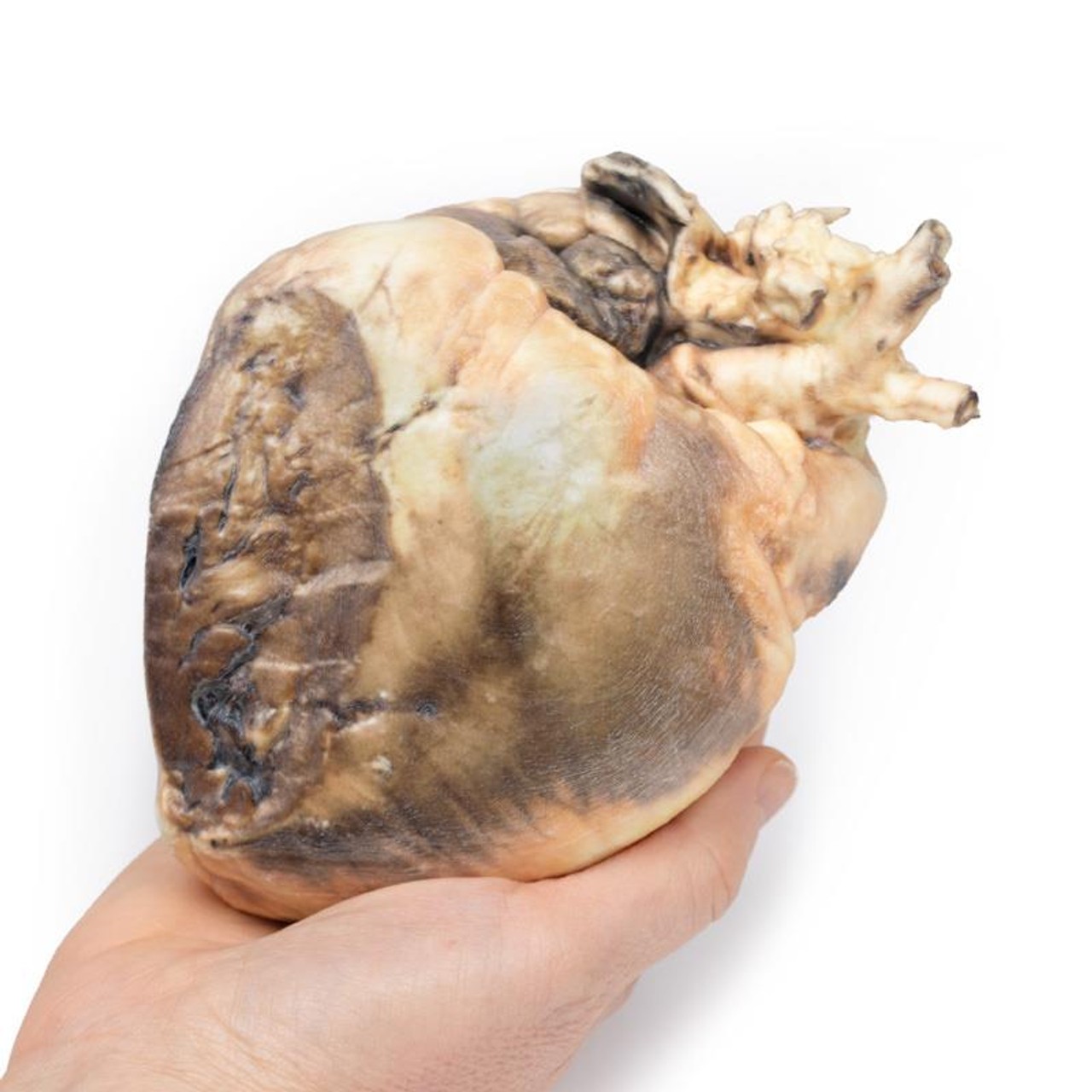
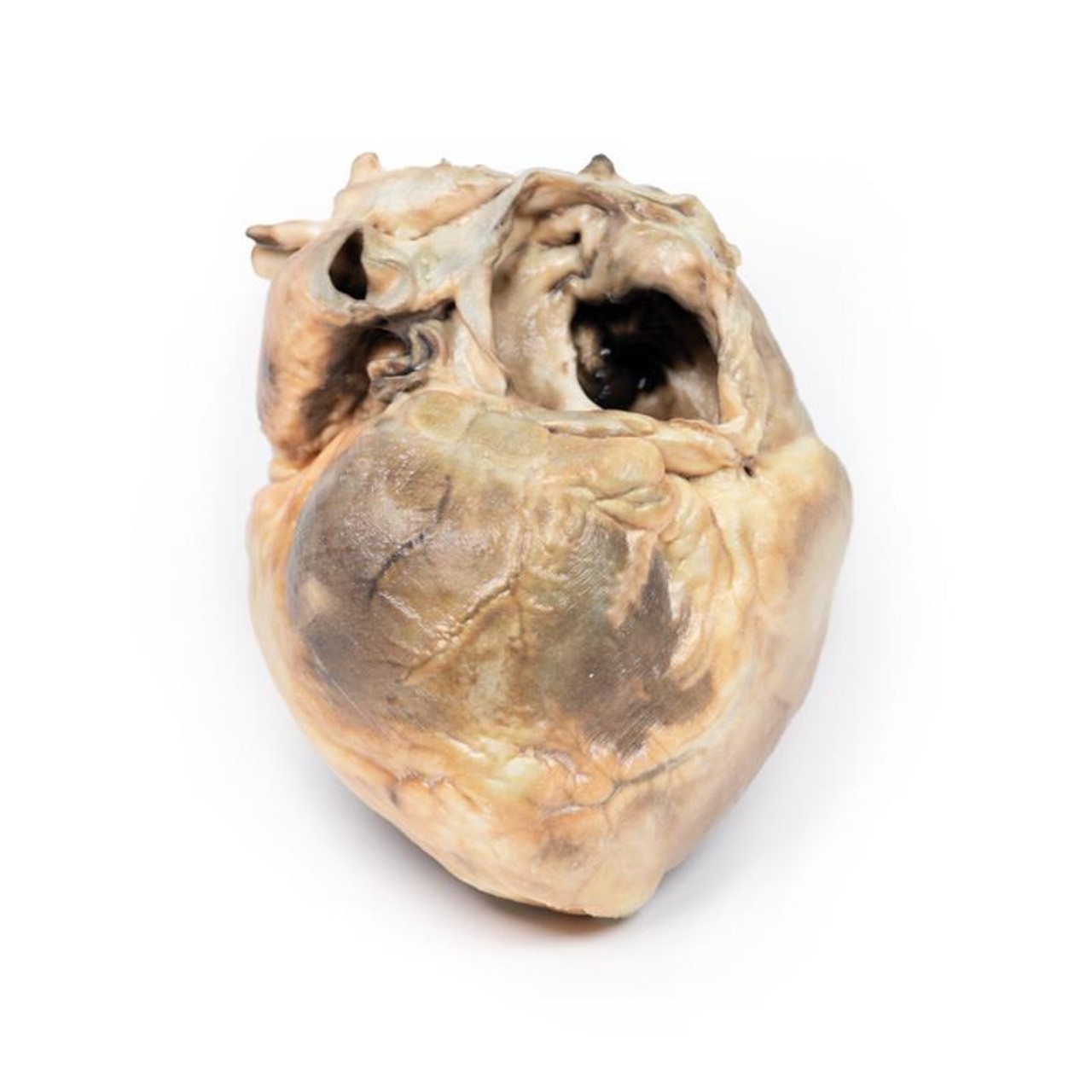
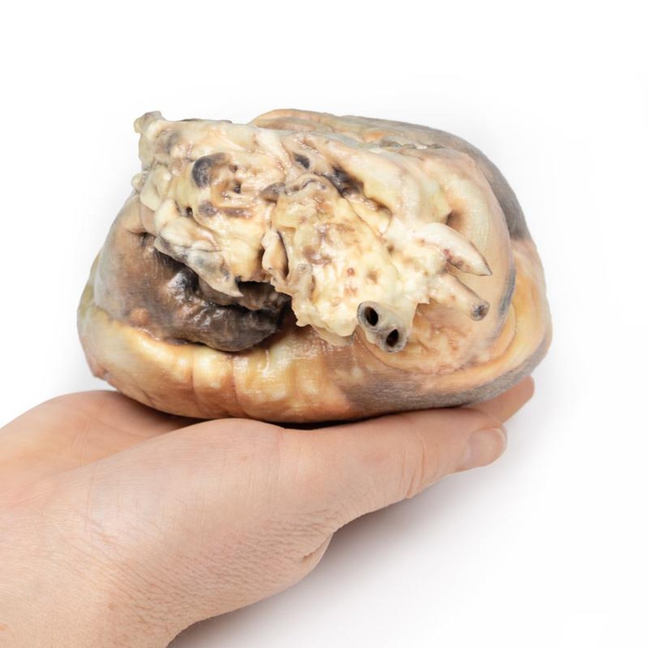
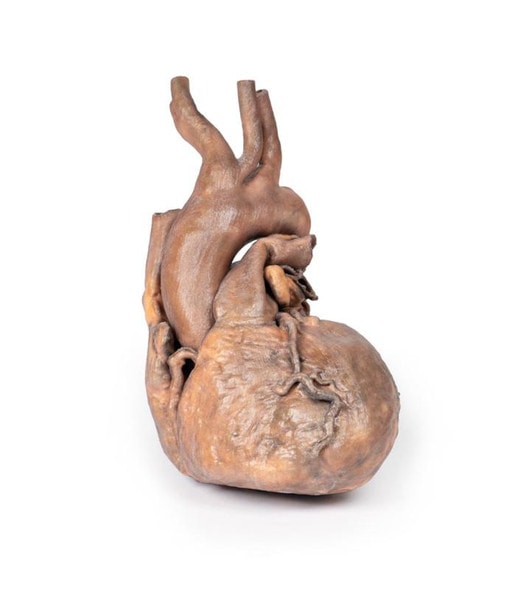
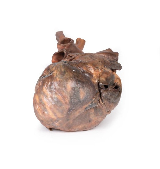
The heart is viewed from the left side. The left atrium has been opened to display a large ovoid defect 3.5 cm in greatest diameter in the inter-atrial septum. Only a small posteroinferior crescentic rim of septum remains. The left ventricle is small, and the right ventricle is hypertrophied (see posterior aspect of specimen where part of the right posterolateral wall of the right ventricle has been cut away to demonstrate the thickened wall). The pulmonary artery, seen to the left of the atrial cavities, is greatly enlarged. The smaller vessel seen lying above the cut end of the pulmonary artery is the aortic arch. The cut edge of a lumen 8 mm in diameter immediately below the cut end of the pulmonary artery is the left auricular appendage.
Further Information
Atrial septal defect is usually asymptomatic early in life, even when large. Symptoms may not develop until adult life. The onset of symptoms is due to reversal of the initial left-to-right shunt as a result of increasing right ventricular hypertrophy and pulmonary hypertension. The ensuing right-to-left shunt is associated with cyanosis and dyspnoea, and ultimately leads to congestive cardiac failure.
There are several types of atrial septal defects, including:
Secundum - This is the most common type of ASD and occurs in the middle of the wall between the atria (atrial septum). Primum - This defect occurs in the lower part of the atrial septum and might occur with other congenital heart problems.
Sinus venosus - This rare defect usually occurs in the upper part of the atrial septum and is often associated with other congenital heart problems.
Coronary sinus - In this rare defect, part of the wall between the coronary sinus which is part of the vein system of the heart and the left atrium is missing.
It is not known why all atrial septal defects occur, but some congenital heart defects appear to be familial and sometimes occur with other genetic problems, such as Trisomy 21 (Down's syndrome). Some conditions during pregnancy can increase the risk of having a baby with a heart defect, including acute infections such as Rubella infection; drug, tobacco or alcohol use, or exposure to certain substances (such as cocaine) during the first trimester of pregnancy; and underlying systemic conditions, such as diabetes or systemic lupus erythematosus.
Advantages of 3D Printed Anatomical Models
- 3D printed anatomical models are the most anatomically accurate examples of human anatomy because they are based on real human specimens.
- Avoid the ethical complications and complex handling, storage, and documentation requirements with 3D printed models when compared to human cadaveric specimens.
- 3D printed anatomy models are far less expensive than real human cadaveric specimens.
- Reproducibility and consistency allow for standardization of education and faster availability of models when you need them.
- Customization options are available for specific applications or educational needs. Enlargement, highlighting of specific anatomical structures, cutaway views, and more are just some of the customizations available.
Disadvantages of Human Cadavers
- Access to cadavers can be problematic and ethical complications are hard to avoid. Many countries cannot access cadavers for cultural and religious reasons.
- Human cadavers are costly to procure and require expensive storage facilities and dedicated staff to maintain them. Maintenance of the facility alone is costly.
- The cost to develop a cadaver lab or plastination technique is extremely high. Those funds could purchase hundreds of easy to handle, realistic 3D printed anatomical replicas.
- Wet specimens cannot be used in uncertified labs. Certification is expensive and time-consuming.
- Exposure to preservation fluids and chemicals is known to cause long-term health problems for lab workers and students. 3D printed anatomical replicas are safe to handle without any special equipment.
- Lack of reuse and reproducibility. If a dissection mistake is made, a new specimen has to be used and students have to start all over again.
Disadvantages of Plastinated Specimens
- Like real human cadaveric specimens, plastinated models are extremely expensive.
- Plastinated specimens still require real human samples and pose the same ethical issues as real human cadavers.
- The plastination process is extensive and takes months or longer to complete. 3D printed human anatomical models are available in a fraction of the time.
- Plastinated models, like human cadavers, are one of a kind and can only showcase one presentation of human anatomy.
Advanced 3D Printing Techniques for Superior Results
- Vibrant color offering with 10 million colors
- UV-curable inkjet printing
- High quality 3D printing that can create products that are delicate, extremely precise, and incredibly realistic
- To improve durability of fragile, thin, and delicate arteries, veins or vessels, a clear support material is printed in key areas. This makes the models robust so they can be handled by students easily.

