Description
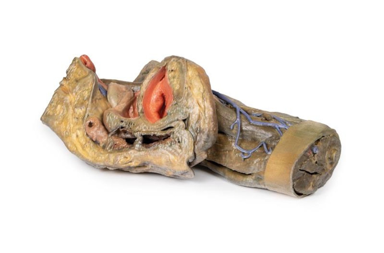
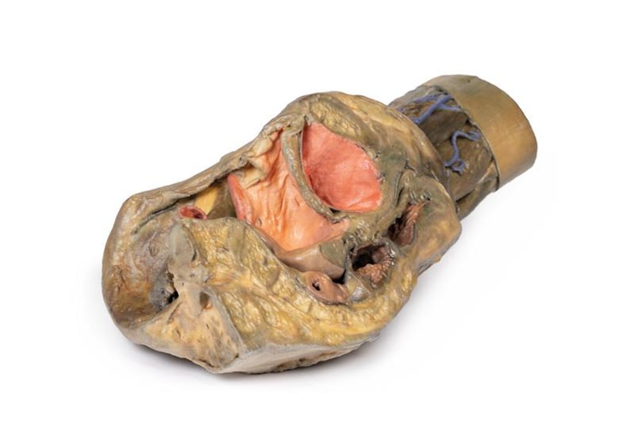
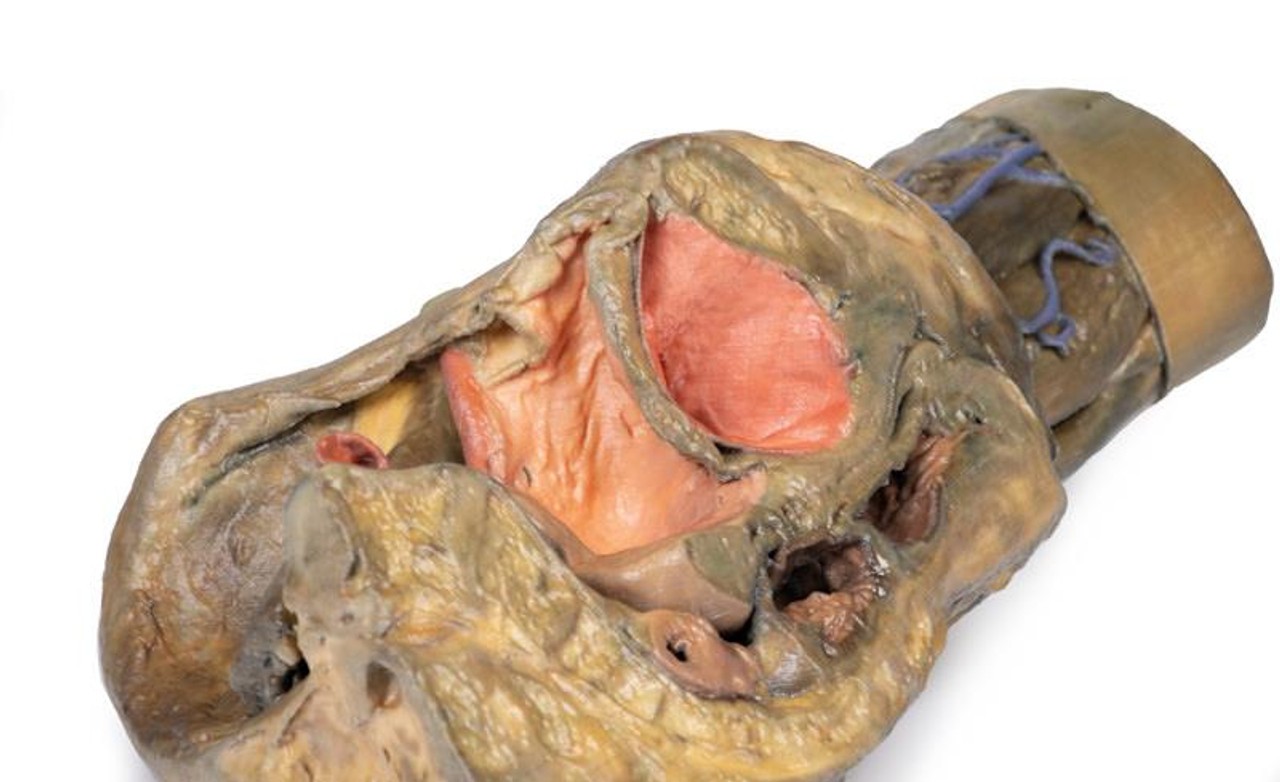
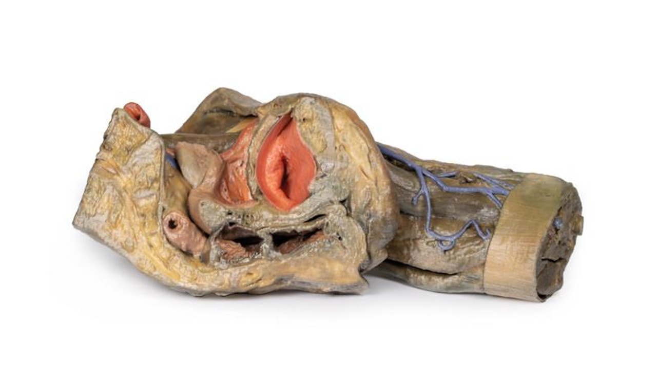
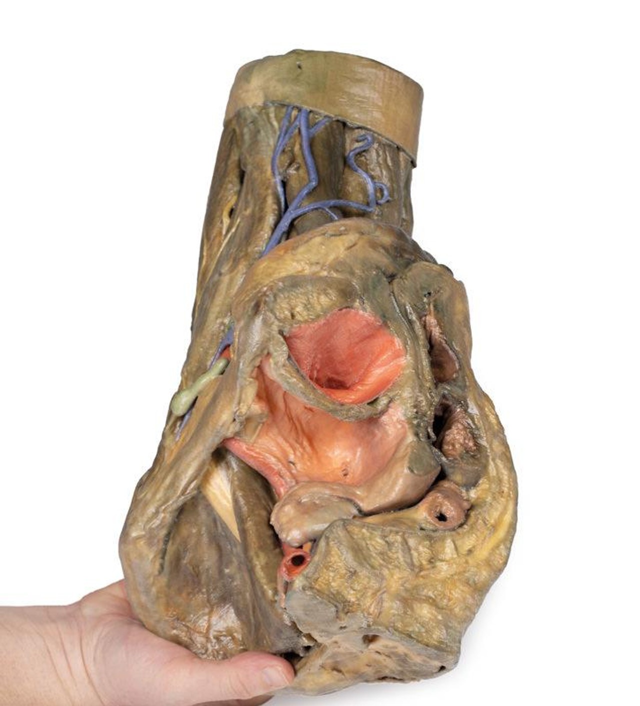
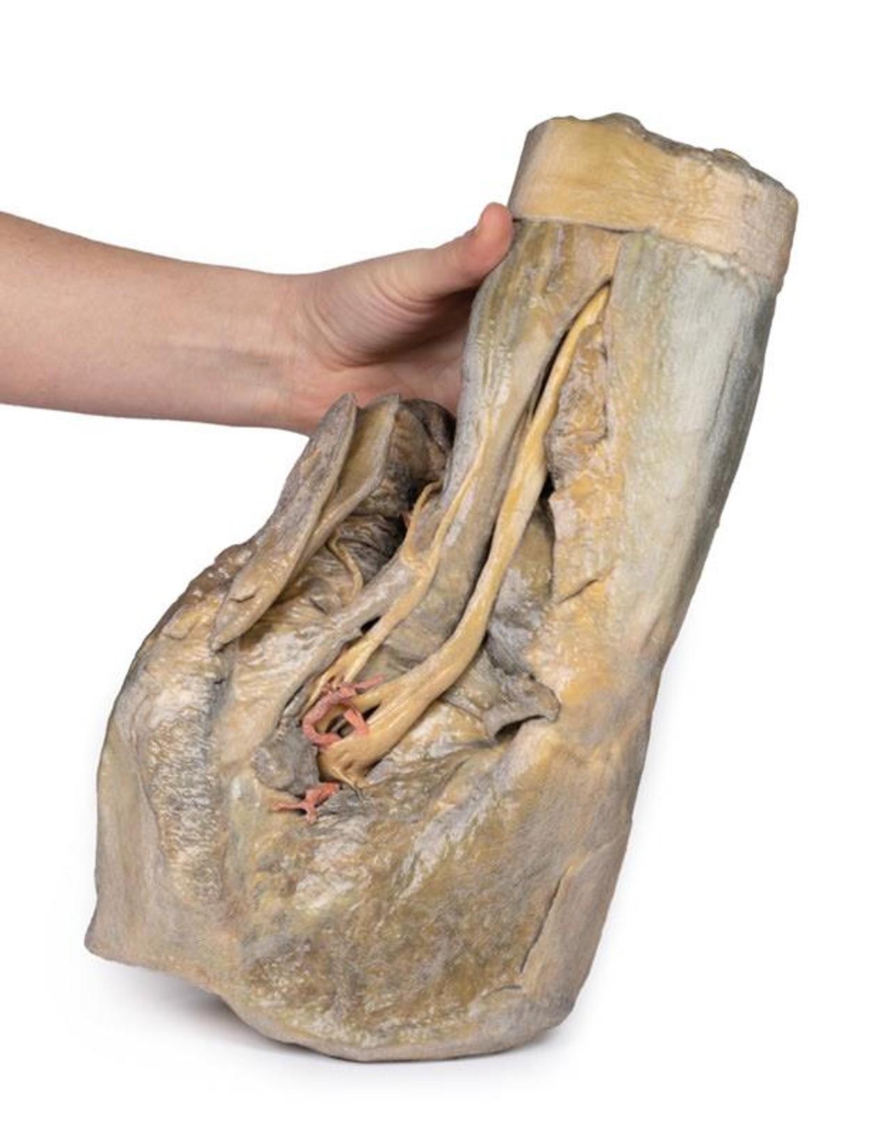
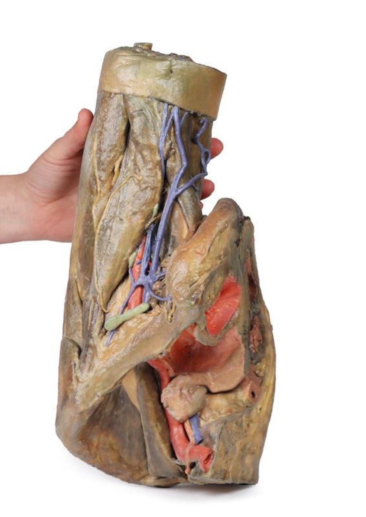
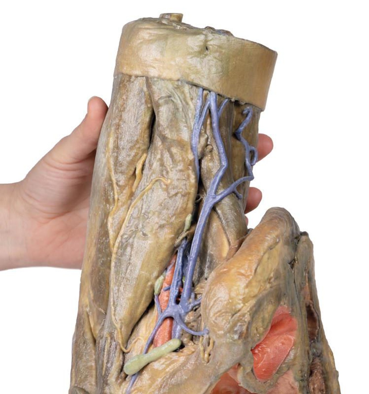
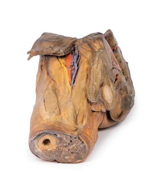
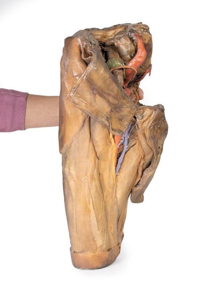
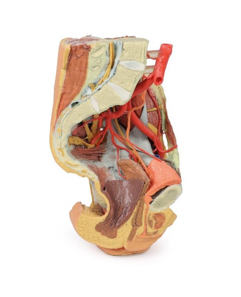
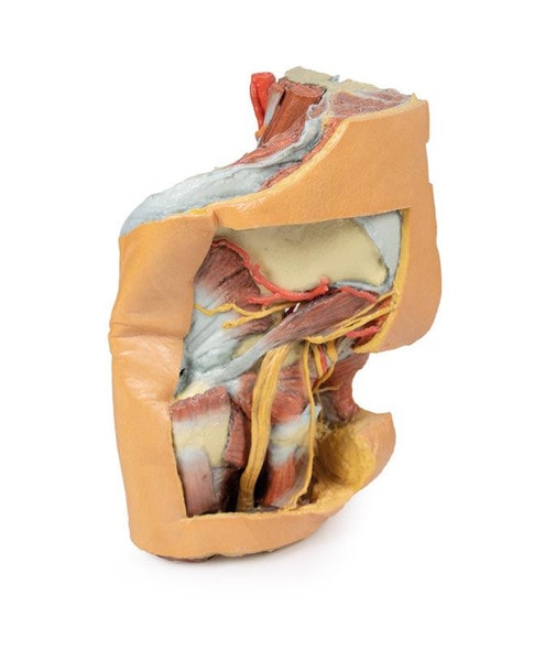
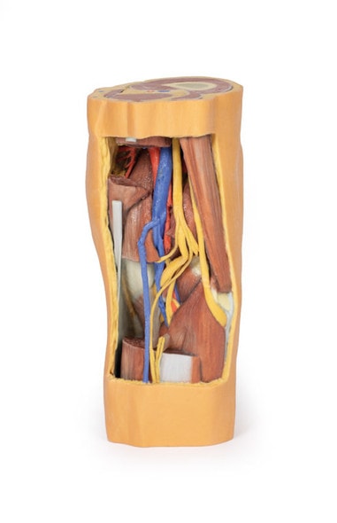
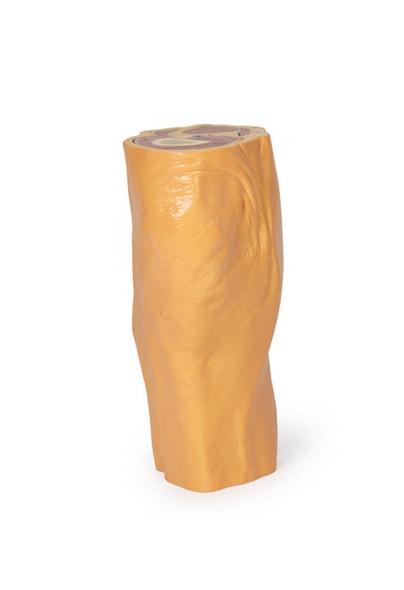
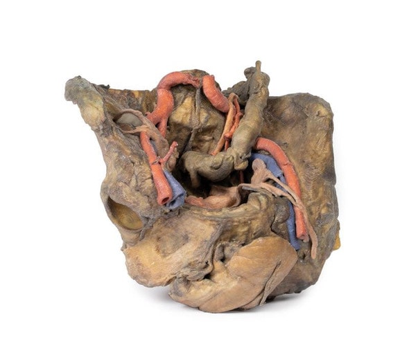
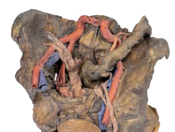
This 3D model preserves a left pelvis divided at the midsagittal plane, and the proximal thigh to approximately the midthigh.
In the midsagittal section, the urinary bladder, uterus and vagina, and rectum can be seen in sequence between the pubic symphysis (anteriorly) and the sacrum (posteriorly). The retention of the peritoneum draped across the superior surface of these organs allows for view of the vesicouterine and rectouterine pouches. The reflection of peritoneum off the uterus forms the broad ligament, with the uterine tube, fimbriae, and closely associated left ovary in position near the pelvic brim. Lateral to the true pelvis contents the common and external iliac arteries can be viewed passing towards the subinguinal space between the common iliac vein and the psoas major muscle. The descending course of the ureter can be traced across these vessels, and the femoral nerve is visible between the psoas major and iliacus muscles.
The superficial fascia has been removed across the entire thigh to the lateral margin of the perineum and near the inferior sectioning of the model itself. Anteriorly, the femoral triangle region has been dissected to expose the content as well as the horizontal group of inguinal lymph nodes immediately inferior to the inguinal ligament. Medially, the femoral vein receives drainage through the great saphenous vein and regional veins (including the superficial circumflex iliac, the superficial external pudendal, and the deep pudendal veins). The femoral artery can be seen immediately lateral to the vein, with parts of the femoral nerve descending just lateral to the artery and near the tendon of the iliopsoas muscle. Although somewhat disturbed by dissection, the anterior cutaneous nerves of the thigh and a small part of the lateral cutaneous nerve of the thigh can be seen on the superficial aspect of the sartorius muscle.
Posteriorly, the gluteal region has been dissected with removal of the gluteus maximus to expose the underlying gluteal muscles, with reflection of the piriformis muscle revealing the neurovascular structures in the region. The sciatic nerve can be seen forming through contributions by the tibial and common peroneal nerves around the preserved portions of the superior and inferior gluteal arteries. Medially the posterior cutaneous nerve of the thigh runs in parallel with the sciatic nerve, with both resting on the obturator internus tendon and gemelli muscles before descending into the thigh on top of the quadratus femoris and common hamstring origin, respectively. Deep to the sacrotuberous ligament the course of the internal pudendal artery and pudendal nerve can be followed towards the ischioanal fossa, where the internal pudendal artery arcs anteriorly and the inferior rectal branch of the pudendal nerve can be seen reaching the pelvic diaphragm and external anal sphincter muscle.
Advantages of 3D Printed Anatomical Models
- 3D printed anatomical models are the most anatomically accurate examples of human anatomy because they are based on real human specimens.
- Avoid the ethical complications and complex handling, storage, and documentation requirements with 3D printed models when compared to human cadaveric specimens.
- 3D printed anatomy models are far less expensive than real human cadaveric specimens.
- Reproducibility and consistency allow for standardization of education and faster availability of models when you need them.
- Customization options are available for specific applications or educational needs. Enlargement, highlighting of specific anatomical structures, cutaway views, and more are just some of the customizations available.
Disadvantages of Human Cadavers
- Access to cadavers can be problematic and ethical complications are hard to avoid. Many countries cannot access cadavers for cultural and religious reasons.
- Human cadavers are costly to procure and require expensive storage facilities and dedicated staff to maintain them. Maintenance of the facility alone is costly.
- The cost to develop a cadaver lab or plastination technique is extremely high. Those funds could purchase hundreds of easy to handle, realistic 3D printed anatomical replicas.
- Wet specimens cannot be used in uncertified labs. Certification is expensive and time-consuming.
- Exposure to preservation fluids and chemicals is known to cause long-term health problems for lab workers and students. 3D printed anatomical replicas are safe to handle without any special equipment.
- Lack of reuse and reproducibility. If a dissection mistake is made, a new specimen has to be used and students have to start all over again.
Disadvantages of Plastinated Specimens
- Like real human cadaveric specimens, plastinated models are extremely expensive.
- Plastinated specimens still require real human samples and pose the same ethical issues as real human cadavers.
- The plastination process is extensive and takes months or longer to complete. 3D printed human anatomical models are available in a fraction of the time.
- Plastinated models, like human cadavers, are one of a kind and can only showcase one presentation of human anatomy.
Advanced 3D Printing Techniques for Superior Results
- Vibrant color offering with 10 million colors
- UV-curable inkjet printing
- High quality 3D printing that can create products that are delicate, extremely precise, and incredibly realistic
- To improve durability of fragile, thin, and delicate arteries, veins or vessels, a clear support material is printed in key areas. This makes the models robust so they can be handled by students easily.

