- Home
- Anatomy Models
- Vascular System Anatomy Models
- 3D Printed Hydatid Disease Affecting the Heart and Aorta
Description
Developed from real patient case study specimens, the 3D printed anatomy model pathology series introduces an unmatched level of realism in human anatomy models. Each 3D printed anatomy model is a high-fidelity replica of a human cadaveric specimen, focusing on the key morbidity presentations that led to the deceasement of the patient. With advances in 3D printing materials and techniques, these stories can come to life in an ethical, consistently reproduceable, and easy to handle format. Ideal for the most advanced anatomical and pathological study, and backed by authentic case study details, students, instructors, and experts alike will discover a new level of anatomical study with the 3D printed anatomy model pathology series.
Clinical History
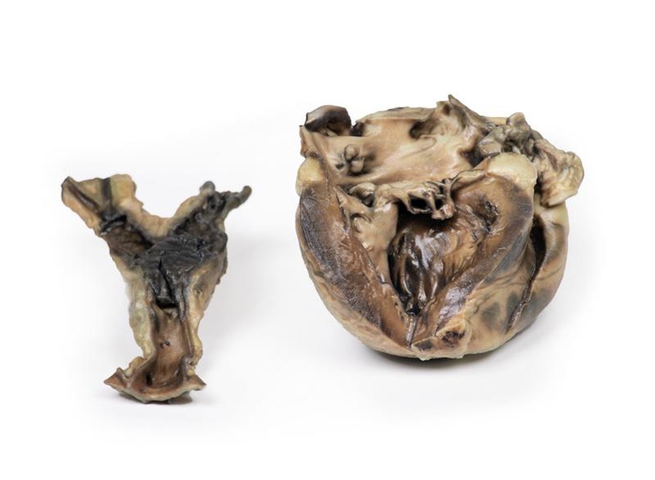
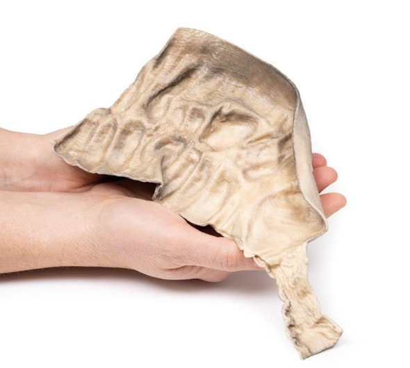
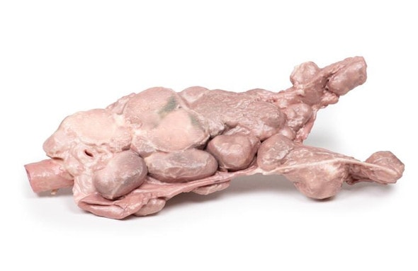
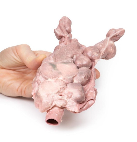
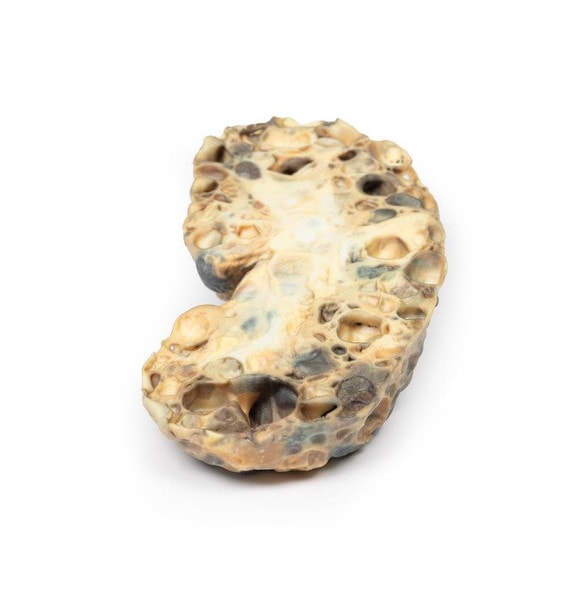
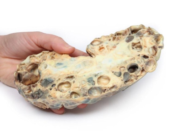
This 11-year-old female had an 18-month history of hydatid disease (see below). In total, 17 cysts were removed from the child's brain at craniotomy on three occasions, and subsequently cysts were found in the kidneys, mesentery, and abdominal aorta at its bifurcation. X-ray of the heart showed a calcified cyst, and the patient was referred to a tertiary hospital for its removal. The patient deteriorated and died following open-heart surgery during which a dead hydatid cyst was found in the left ventricle.
Pathology
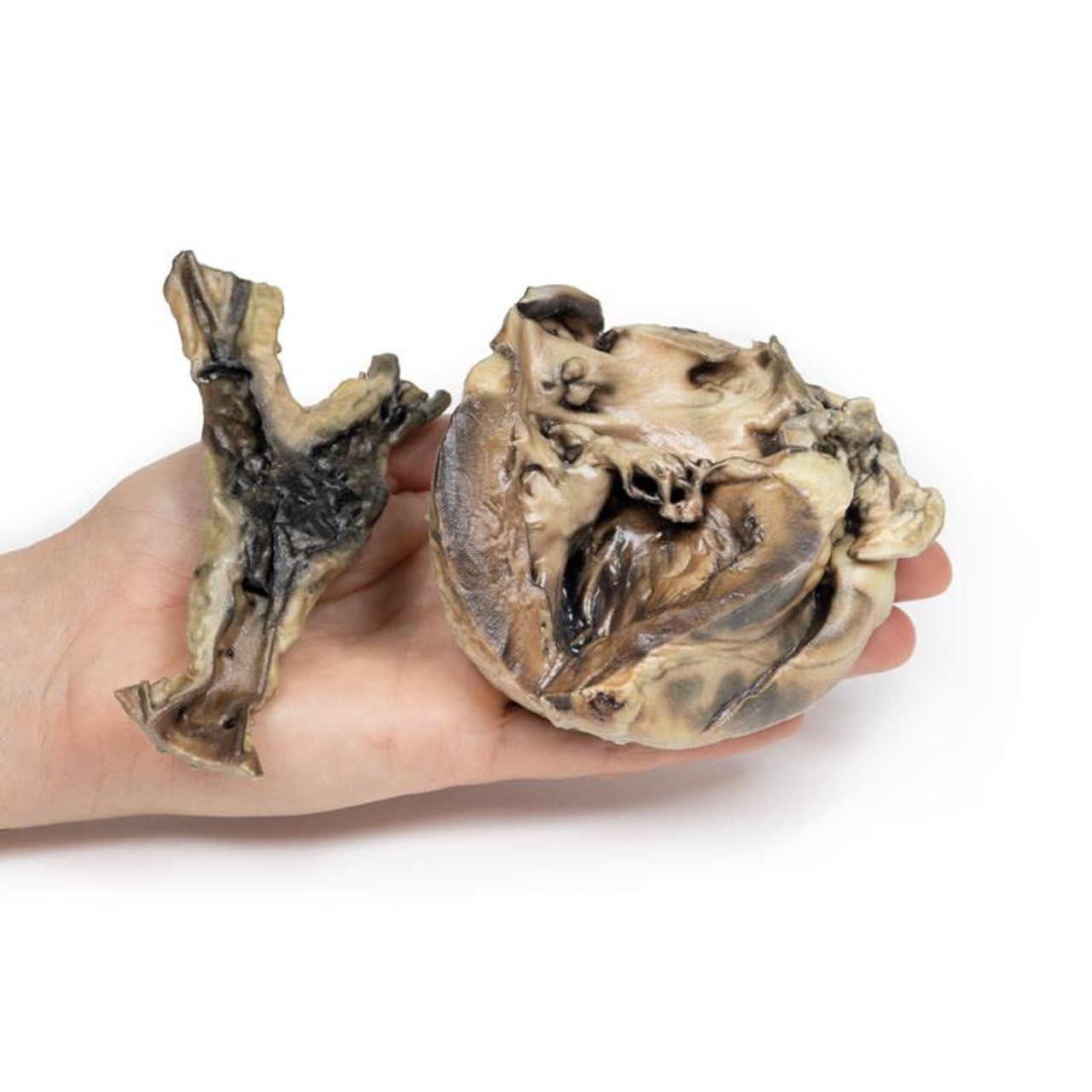
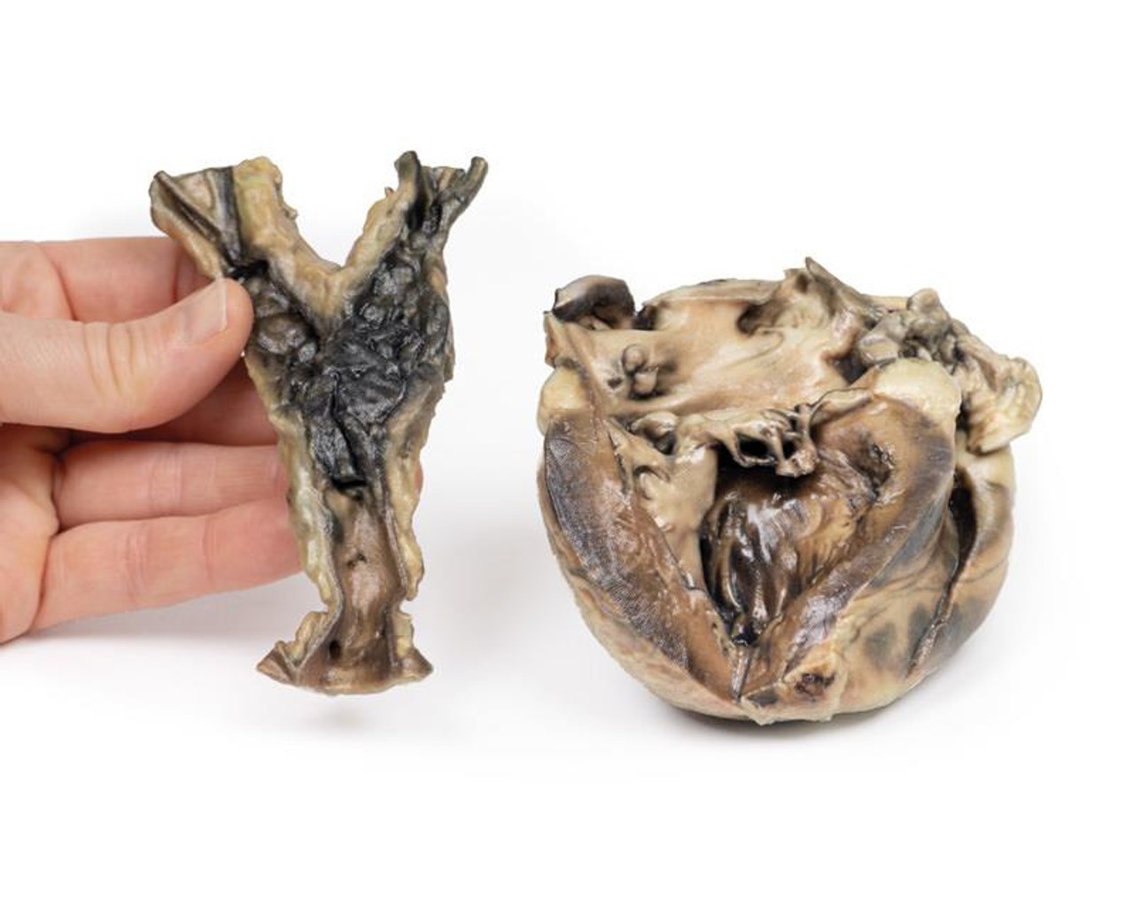
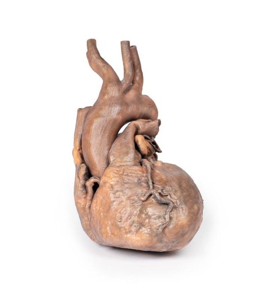
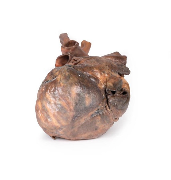
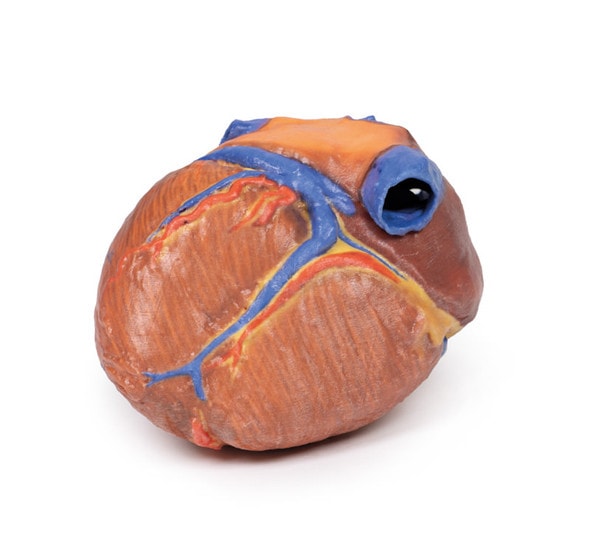
The specimens are of the heart, with the left ventricle being laid open, and of the aorta at its common iliac bifurcation. The aorta shows some atheromatous depositions in the upper portion. There is a large mass of antemortem clot at the point of iliac bifurcation with extension down both common iliac arteries.
The heart shows hypertrophy of the left ventricular wall, and an abnormal communication between the left ventricle and atrium running through the posterior cusp of the mitral valve via the papillary muscle into the left ventricular cavity. This channel is surrounded by thickened fibrous-looking tissue. The posterior cusp of the valve has been split. Sutured surgical incisions are visible on the posterior aspect of the specimen and the ventricular wall and in the left atrial appendage. Hydatid cysts occupy the abdominal aorta at its bifurcation, and the channel joining the left ventricle and left atrium.
Histology demonstrated cysts within the aorta wall comprised of 3 layers: an outermost pericyst fibrous layer; a middle ectocyst layer that was laminated, hyaline and acellular; and the inner endocyst in the germinative layer, consisting of daughter cysts and brood capsules with scolices. A focal granulomatous palisading reaction was also present within the aorta wall.
Further Information
Hydatid cyst is a human parasitic disease caused by the larval stage of the cestode tapeworm Echinococcus granulosus, which infests the gut of dogs its definitive hosts. Human beings may serve as incidental hosts by the ingestion of ova in vegetables or water contaminated with dog feces. Humans become infected by the ingestion of eggs passed in dog feces. Oncospheres released from the eggs penetrate the intestinal mucosa and, via the portal system, lodge in the liver, lungs, muscle or other organs, where the hydatid cysts form.
Hydatid disease is endemic in cattle-raising areas of the world, notably in the Mediterranean countries, the Middle East, South America, Australia, and New Zealand. Although no body part can be spared from hydatid cysts, they mostly affect the liver and lungs. Cardiac involvement is much rarer, yet potentially fatal condition and comprises 0.5–2% of all hydatid cases. Cardiac complications and presentation vary depends on the location, size and integrity of the cyst(s). The myocardium of the left ventricle more frequently involved. Pericardial involvement occurs mostly in multifocal cardiac echinococcosis. Growth of the cyst leads them being pushed toward a weaker side of the cardiac wall, either the epicardium or the endocardium. LV HCs are usually located subepicardially, therefore rarely rupture into the pericardial space. However, if rupture happens, it may be silent or it may cause acute pericardial tamponade, constrictive pericarditis or secondary pericardial cysts[1].
Although E. granulosus is still found in sheep and rural dogs in Australia, the prevalence of transmission is less common than it was. The marked reduction in prevalence in rural domestic dogs, and also sheep, is the result of the highly effective cestocidal drug, praziquantel, being included in
Readily available, cheap, generic, all-wormers for dogs and the development of inexpensive commercial dry dog food[2].
References
1. Oraha et al. Ann Med Surg (Lond). 2018
2. Jenkins et al. Int J Parasitol Parasites Wildl. 2019
Advantages of 3D Printed Anatomical Models
- 3D printed anatomical models are the most anatomically accurate examples of human anatomy because they are based on real human specimens.
- Avoid the ethical complications and complex handling, storage, and documentation requirements with 3D printed models when compared to human cadaveric specimens.
- 3D printed anatomy models are far less expensive than real human cadaveric specimens.
- Reproducibility and consistency allow for standardization of education and faster availability of models when you need them.
- Customization options are available for specific applications or educational needs. Enlargement, highlighting of specific anatomical structures, cutaway views, and more are just some of the customizations available.
Disadvantages of Human Cadavers
- Access to cadavers can be problematic and ethical complications are hard to avoid. Many countries cannot access cadavers for cultural and religious reasons.
- Human cadavers are costly to procure and require expensive storage facilities and dedicated staff to maintain them. Maintenance of the facility alone is costly.
- The cost to develop a cadaver lab or plastination technique is extremely high. Those funds could purchase hundreds of easy to handle, realistic 3D printed anatomical replicas.
- Wet specimens cannot be used in uncertified labs. Certification is expensive and time-consuming.
- Exposure to preservation fluids and chemicals is known to cause long-term health problems for lab workers and students. 3D printed anatomical replicas are safe to handle without any special equipment.
- Lack of reuse and reproducibility. If a dissection mistake is made, a new specimen has to be used and students have to start all over again.
Disadvantages of Plastinated Specimens
- Like real human cadaveric specimens, plastinated models are extremely expensive.
- Plastinated specimens still require real human samples and pose the same ethical issues as real human cadavers.
- The plastination process is extensive and takes months or longer to complete. 3D printed human anatomical models are available in a fraction of the time.
- Plastinated models, like human cadavers, are one of a kind and can only showcase one presentation of human anatomy.
Advanced 3D Printing Techniques for Superior Results
- Vibrant color offering with 10 million colors
- UV-curable inkjet printing
- High quality 3D printing that can create products that are delicate, extremely precise, and incredibly realistic
- To improve durability of fragile, thin, and delicate arteries, veins or vessels, a clear support material is printed in key areas. This makes the models robust so they can be handled by students easily.

