- Home
- Anatomy Models
- Abdomen Anatomy Models
- 3D Printed Mesenteric Metastases from Cutaneous Malignant Melanoma
Description
Developed from real patient case study specimens, the 3D printed anatomy model pathology series introduces an unmatched level of realism in human anatomy models. Each 3D printed anatomy model is a high-fidelity replica of a human cadaveric specimen, focusing on the key morbidity presentations that led to the deceasement of the patient. With advances in 3D printing materials and techniques, these stories can come to life in an ethical, consistently reproduceable, and easy to handle format. Ideal for the most advanced anatomical and pathological study, and backed by authentic case study details, students, instructors, and experts alike will discover a new level of anatomical study with the 3D printed anatomy model pathology series.
Clinical History
A 44-year-old man had a skin lesion on his back that grew slowly. At presentation at A&E several years later, he complained of bone pain, and had hepatomegaly and a pleural effusion. He died shortly afterwards.
Pathology
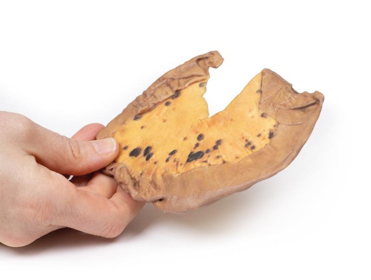
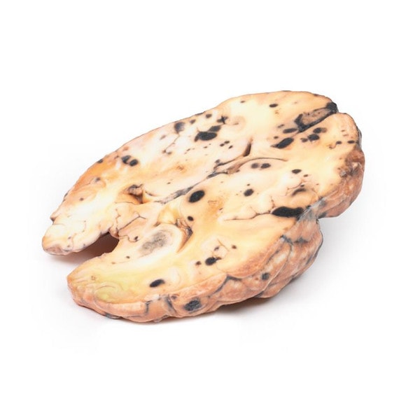
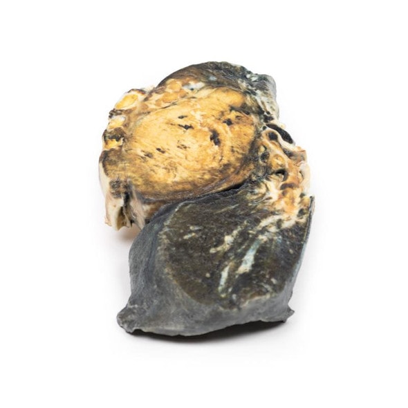
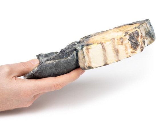
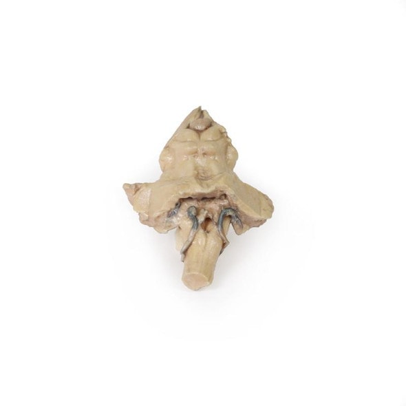
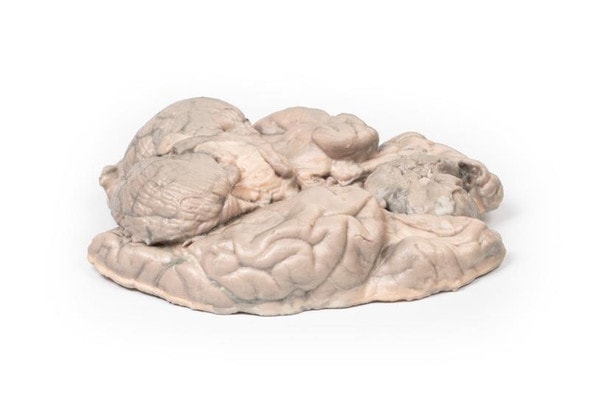
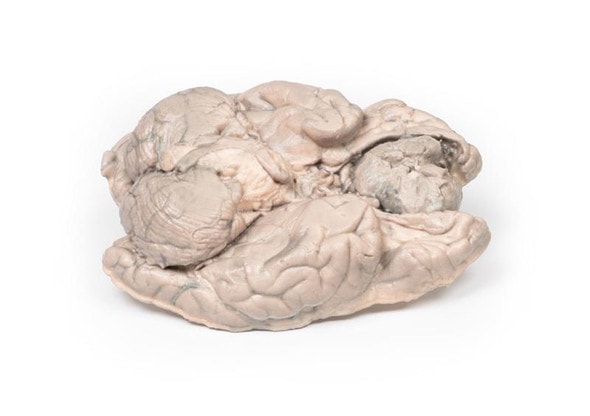
The specimen is a loop of small intestine mounted to display the mesentery, which contains numerous small dark brown, circumscribed nodules varying from pin head size to approximately 1 cm in diameter. Histology confirmed the diagnosis of metastatic melanoma.
Further Information
The most common form of melanoma is cutaneous melanoma, which develops from the pigment-producing cells known as melanocytes. In women, they most commonly occur on the legs, while in men they most commonly occur on the back. About 25% of melanomas develop from moles. Changes in a mole that can indicate melanoma include an increase in size, irregular edges, change in color, itchiness or skin ulceration.
Skin melanoma is associated with exposure to UV radiation in sunlight or tanning beds. Other risk factors for developing melanoma include fair complexion, presence of large number of melanocytic naevi (moles), severe sunburn as a child, and immunosuppression. It accounts for around 5% of all skin cancer diagnosis but has the highest mortality rate of all skin cancers. Melanomas typically occur in sun exposed areas as a pigmented lesion with irregular borders, variegated color, an asymmetrical shape and which evolves with time.
There are multiple mutations common in melanoma. Loss of cell cycle control gene from mutation in CDKN2A gene. Mutations in pro-growth signaling pathways, such as BRAF and PI3K mutations, are seen frequently in melanomas as well as mutations that activate telomerase, such as the TERT gene. Recognition that melanoma antigens activate host immune responses has led to promising immunotherapy, which enhances host T-cell identifying of these antigens.
The most common sites for metastasis of melanoma are the lungs, liver, brain and bone as well as regional lymph nodes, and is highly dependent on the site of the primary tumor. Metastatic melanoma involving the gastrointestinal tract may present with anemia, overt bleeding, pain, obstruction, or intussusception. The jejunum and ileum are the most commonly involved sites, followed by the colon, rectum, and stomach. Surgery has usually been reserved for patients with the above complications.
The probability of metastatic spread from skin melanoma depends on the stage of the primary tumor, which is based on tumor depth, mitotic activity and ulceration of the skin as well as node and solid organ involvement. Diagnosis of melanoma is made with excisional biopsy. Investigation for bone metastasis is done using blood test (raised Alkaline phosphatase, calcium and LDH), and radiological investigations most commonly X-ray and CT but MRI and PET scans may also be used. Treatment depends on the stage or the tumor as well as the genetic and immune profiles of the melanoma. Treatment usually involves surgical resection, chemotherapy, targeted therapies (e.g. BRAF inhibitors), immunotherapy , radiotherapy or more commonly a combination of treatments.
Advantages of 3D Printed Anatomical Models
- 3D printed anatomical models are the most anatomically accurate examples of human anatomy because they are based on real human specimens.
- Avoid the ethical complications and complex handling, storage, and documentation requirements with 3D printed models when compared to human cadaveric specimens.
- 3D printed anatomy models are far less expensive than real human cadaveric specimens.
- Reproducibility and consistency allow for standardization of education and faster availability of models when you need them.
- Customization options are available for specific applications or educational needs. Enlargement, highlighting of specific anatomical structures, cutaway views, and more are just some of the customizations available.
Disadvantages of Human Cadavers
- Access to cadavers can be problematic and ethical complications are hard to avoid. Many countries cannot access cadavers for cultural and religious reasons.
- Human cadavers are costly to procure and require expensive storage facilities and dedicated staff to maintain them. Maintenance of the facility alone is costly.
- The cost to develop a cadaver lab or plastination technique is extremely high. Those funds could purchase hundreds of easy to handle, realistic 3D printed anatomical replicas.
- Wet specimens cannot be used in uncertified labs. Certification is expensive and time-consuming.
- Exposure to preservation fluids and chemicals is known to cause long-term health problems for lab workers and students. 3D printed anatomical replicas are safe to handle without any special equipment.
- Lack of reuse and reproducibility. If a dissection mistake is made, a new specimen has to be used and students have to start all over again.
Disadvantages of Plastinated Specimens
- Like real human cadaveric specimens, plastinated models are extremely expensive.
- Plastinated specimens still require real human samples and pose the same ethical issues as real human cadavers.
- The plastination process is extensive and takes months or longer to complete. 3D printed human anatomical models are available in a fraction of the time.
- Plastinated models, like human cadavers, are one of a kind and can only showcase one presentation of human anatomy.
Advanced 3D Printing Techniques for Superior Results
- Vibrant color offering with 10 million colors
- UV-curable inkjet printing
- High quality 3D printing that can create products that are delicate, extremely precise, and incredibly realistic
- To improve durability of fragile, thin, and delicate arteries, veins or vessels, a clear support material is printed in key areas. This makes the models robust so they can be handled by students easily.


