Description
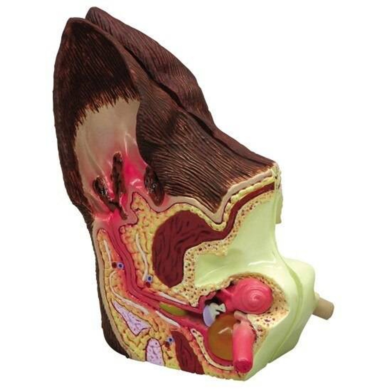
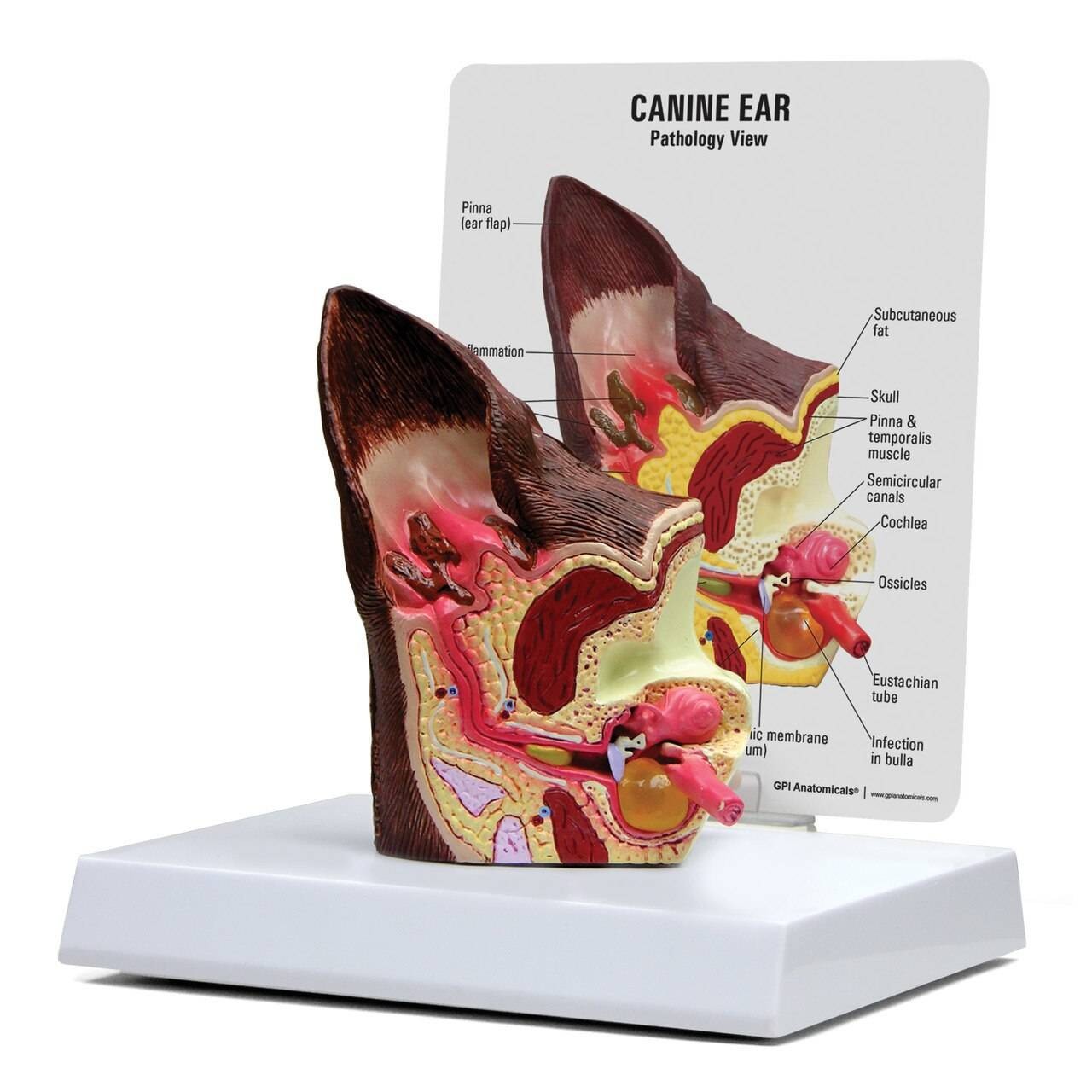
The Canine Ear Model shows normal anatomy and structures on one side and pathologies on the other. The normal side displays the cochlea, auditory ossicles, auditory tube, tympanic bulla, middle ear cavity, tympanic membrane, horizontal canal, vertical canal, auricular cartilage, pinna and temporalis muscle. The abnormal side illustrates inflamed inner ear structures, inflammatory exudate in tympanic bulla, ear canal with partial occlusion from cellular hyperplasia, inflammatory exudate and an inflamed outer ear. Includes key card. The model is a fantastic addition to any veterinary setting, especially for advanced study and patient consultation.
The Canine Ear Model measures: 3 x 4 x 1-1/2. Made by GPI Anatomicals.

