Description
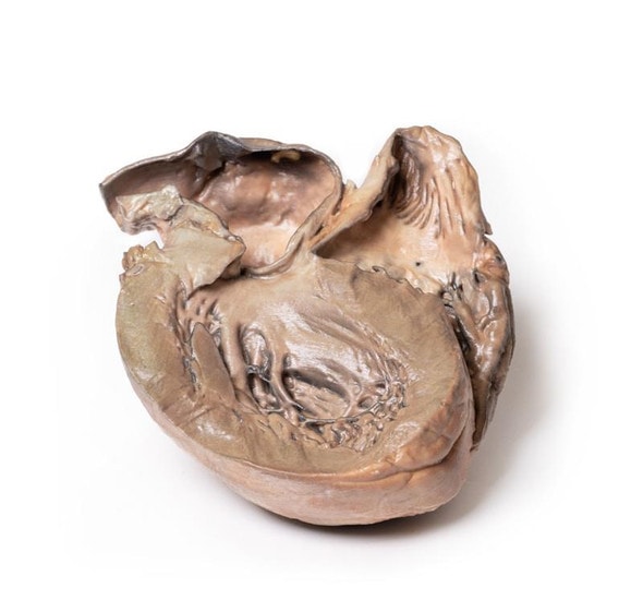
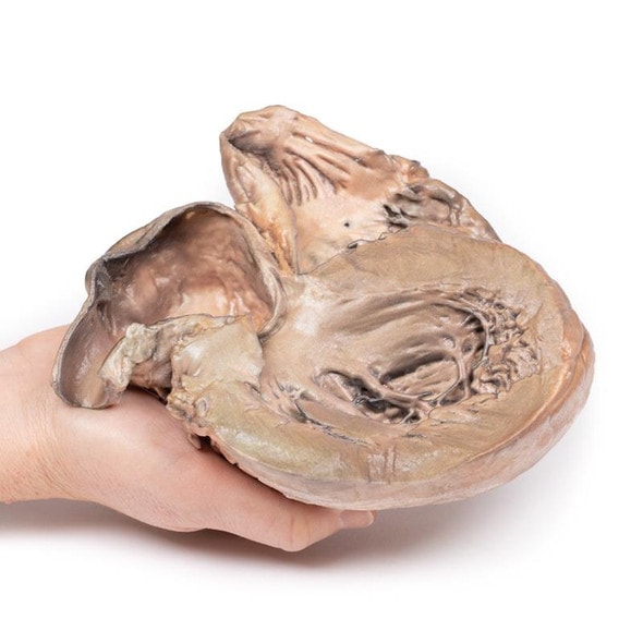
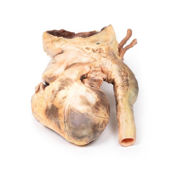
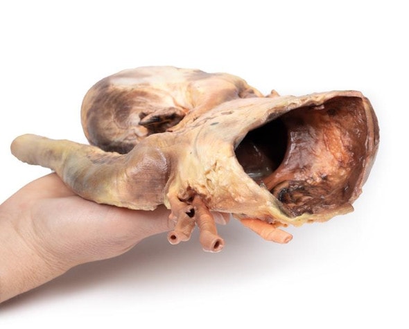
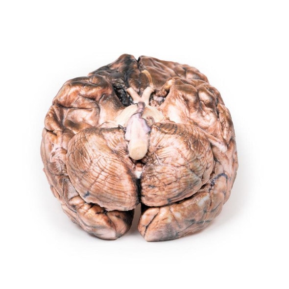
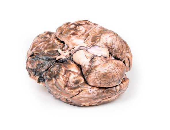
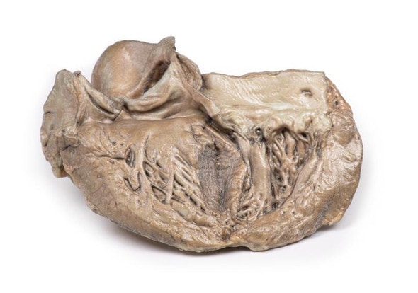
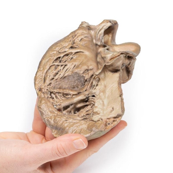
Developed from real patient case study specimens, the 3D printed anatomy model pathology series introduces an unmatched level of realism in human anatomy models. Each 3D printed anatomy model is a high-fidelity replica of a human cadaveric specimen, focusing on the key morbidity presentations that led to the deceasement of the patient. With advances in 3D printing materials and techniques, these stories can come to life in an ethical, consistently reproduceable, and easy to handle format. Ideal for the most advanced anatomical and pathological study, and backed by authentic case study details, students, instructors, and experts alike will discover a new level of anatomical study with the 3D printed anatomy model pathology series.
Clinical History
This 70-year-old man with a past history of mild gastro-oesophageal reflux presented to the Alfred Hospital with a sudden onset of severe upper abdominal pain, which radiated to the left shoulder tip. On examination, he was distressed and hyperventilating, pulse rate 87/min, and a blood pressure 140/90 mm Hg. Abdominal examination revealed board-like rigidity and diminished bowel sounds. At emergency laparotomy no evidence of a ruptured viscus was found; the pancreas appeared normal and an unruptured abdominal aortic aneurism was noted. Endoscopy on the following day showed a ruptured oesophageal ulcer and a Celestin tube was inserted. The patient developed localized infective complications, pulmonary oedema and congestion, and died 19 days after admission.
Pathology
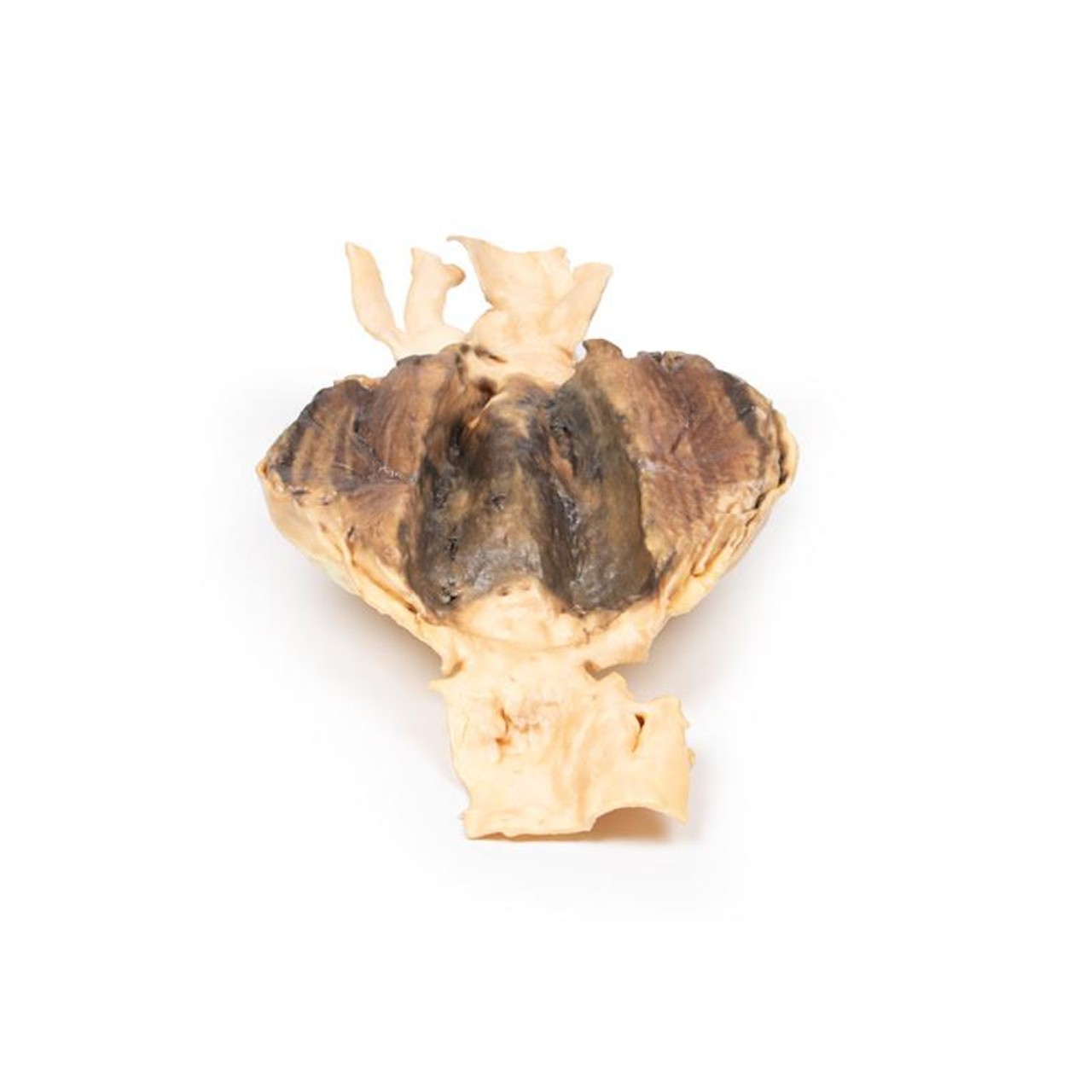
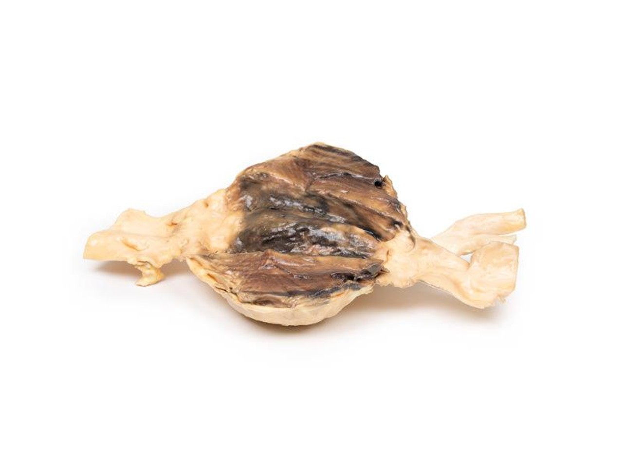
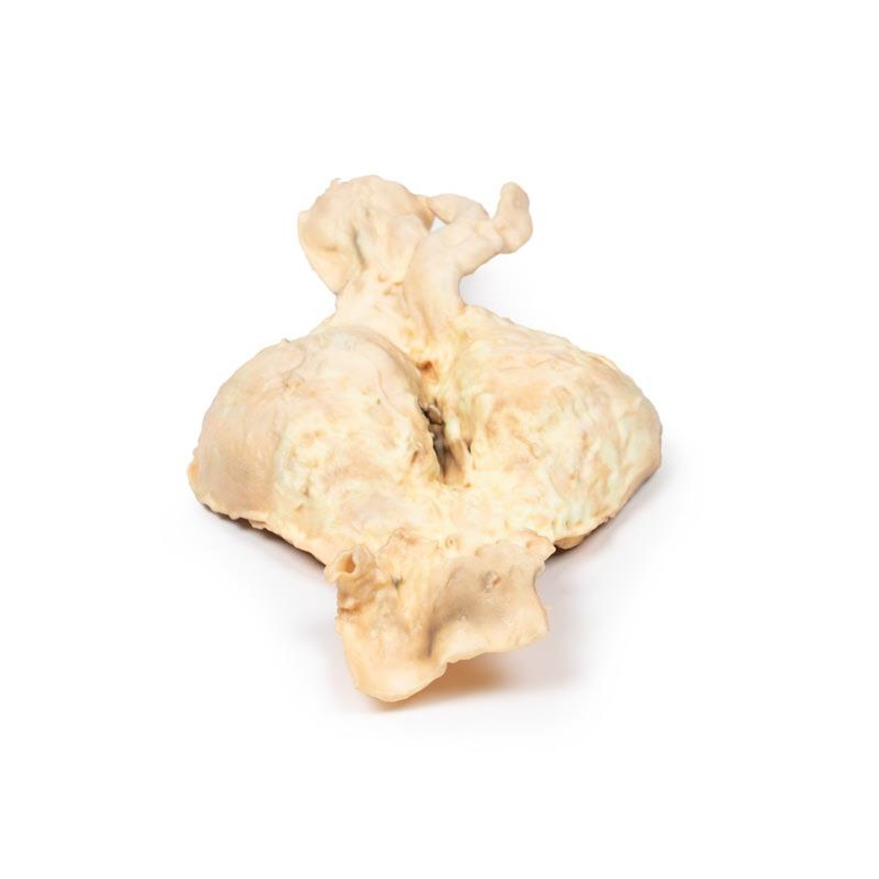
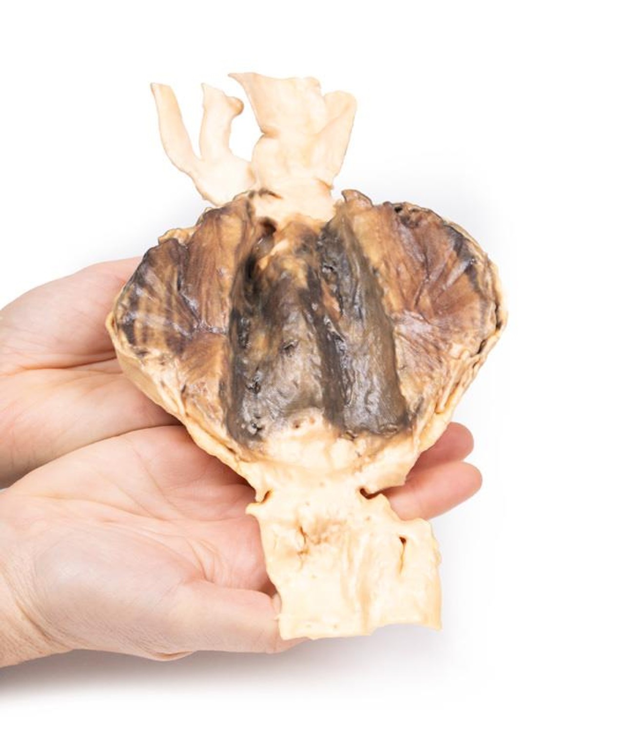
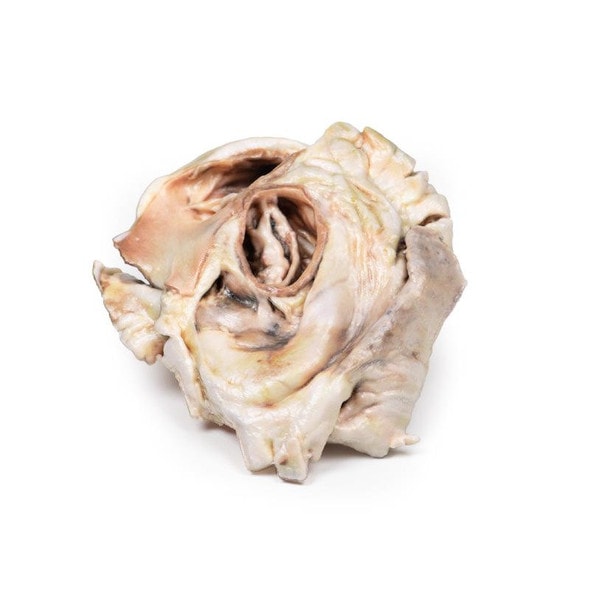
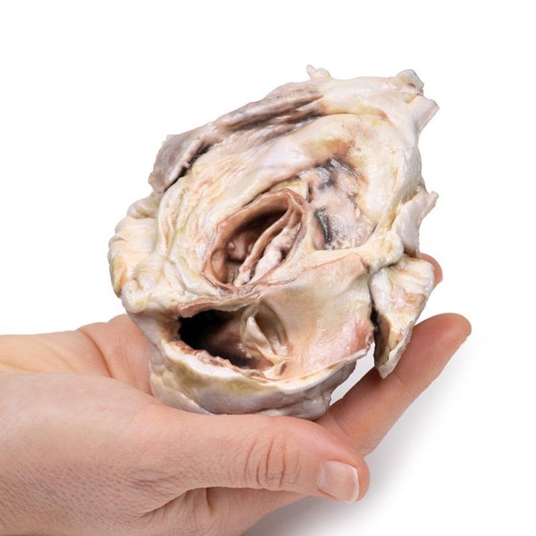
The specimen consists of lower abdominal segment of aorta together with common iliac vessels and proximal portions of the internal and external iliac arteries. A large 10 x 7 cm aneurysm is situated below the origin of the renal arteries extending to the aortic bifurcation. The aneurysm with its severe thinning of the wall of the abdominal aorta is partly lined by a laminated thrombus, indicating the chronicity of the process. There is evidence of a recent thrombus on the luminal surface. There also appears to be some aneurysmal dilatation of the common iliac and (opened) proximal left external iliac artery. The abdominal aorta at the upper end of the specimen shows multiple focally ulcerated atheromatous plaques. There is no evidence of rupture.
Further Information
Abdominal aortic aneurysm (AAA or triple A) represents a localized enlargement of the abdominal aorta (diameter >3 cm or more than 50% larger than normal)[1]. They are usually asymptomatic, except during rupture[1]. Large aneurysms may be palpable on abdominal examination. Occasionally, abdominal-, back-, or leg pain may occur depending on location and size. Rupture may result in pain in the abdomen or back, sudden low blood pressure with loss of consciousness, and often results in death[1]. AAA's occur most commonly in those over 50 years of age, in men, and amongst those with a family history of this disease. Additional risk factors include smoking, high blood pressure, and other heart or blood vessel diseases. They are also found in genetic abnormalities, including Marfan's syndrome and Ehlers-Danlos syndrome. AAAs are the most common form of aortic aneurysm, and about 85% occur below the kidneys.
Advantages of 3D Printed Anatomical Models
- 3D printed anatomical models are the most anatomically accurate examples of human anatomy because they are based on real human specimens.
- Avoid the ethical complications and complex handling, storage, and documentation requirements with 3D printed models when compared to human cadaveric specimens.
- 3D printed anatomy models are far less expensive than real human cadaveric specimens.
- Reproducibility and consistency allow for standardization of education and faster availability of models when you need them.
- Customization options are available for specific applications or educational needs. Enlargement, highlighting of specific anatomical structures, cutaway views, and more are just some of the customizations available.
Disadvantages of Human Cadavers
- Access to cadavers can be problematic and ethical complications are hard to avoid. Many countries cannot access cadavers for cultural and religious reasons.
- Human cadavers are costly to procure and require expensive storage facilities and dedicated staff to maintain them. Maintenance of the facility alone is costly.
- The cost to develop a cadaver lab or plastination technique is extremely high. Those funds could purchase hundreds of easy to handle, realistic 3D printed anatomical replicas.
- Wet specimens cannot be used in uncertified labs. Certification is expensive and time-consuming.
- Exposure to preservation fluids and chemicals is known to cause long-term health problems for lab workers and students. 3D printed anatomical replicas are safe to handle without any special equipment.
- Lack of reuse and reproducibility. If a dissection mistake is made, a new specimen has to be used and students have to start all over again.
Disadvantages of Plastinated Specimens
- Like real human cadaveric specimens, plastinated models are extremely expensive.
- Plastinated specimens still require real human samples and pose the same ethical issues as real human cadavers.
- The plastination process is extensive and takes months or longer to complete. 3D printed human anatomical models are available in a fraction of the time.
- Plastinated models, like human cadavers, are one of a kind and can only showcase one presentation of human anatomy.
Advanced 3D Printing Techniques for Superior Results
- Vibrant color offering with 10 million colors
- UV-curable inkjet printing
- High quality 3D printing that can create products that are delicate, extremely precise, and incredibly realistic
- To improve durability of fragile, thin, and delicate arteries, veins or vessels, a clear support material is printed in key areas. This makes the models robust so they can be handled by students easily.

