Description
Developed from real patient case study specimens, the 3D printed anatomy model pathology series introduces an unmatched level of realism in human anatomy models. Each 3D printed anatomy model is a high-fidelity replica of a human cadaveric specimen, focusing on the key morbidity presentations that led to the deceasement of the patient. With advances in 3D printing materials and techniques, these stories can come to life in an ethical, consistently reproduceable, and easy to handle format. Ideal for the most advanced anatomical and pathological study, and backed by authentic case study details, students, instructors, and experts alike will discover a new level of anatomical study with the 3D printed anatomy model pathology series.
Clinical History
A 61-year old male presents with exertional anginal chest pain and dyspnoea. He has had these symptoms for 6 years with increasing severity. On examination, he is cyanotic and tachycardic with a collapsing pulse. A swelling was noted on the right side of his neck. There was a thrill in his carotid artery. The apex beat was displaced inferolaterally. A loud systolic and diastolic murmur was auscultated in the aortic area. Chest X-rays showed cardiomegaly with a large rounded lesion in the right upper mediastinum continuous with the heart shadow with radiographic evidence of cardiac failure. Blood tests were positive for anti-treponemal antibodies. The patient's condition deteriorated and he died of cardiac failure.
Pathology
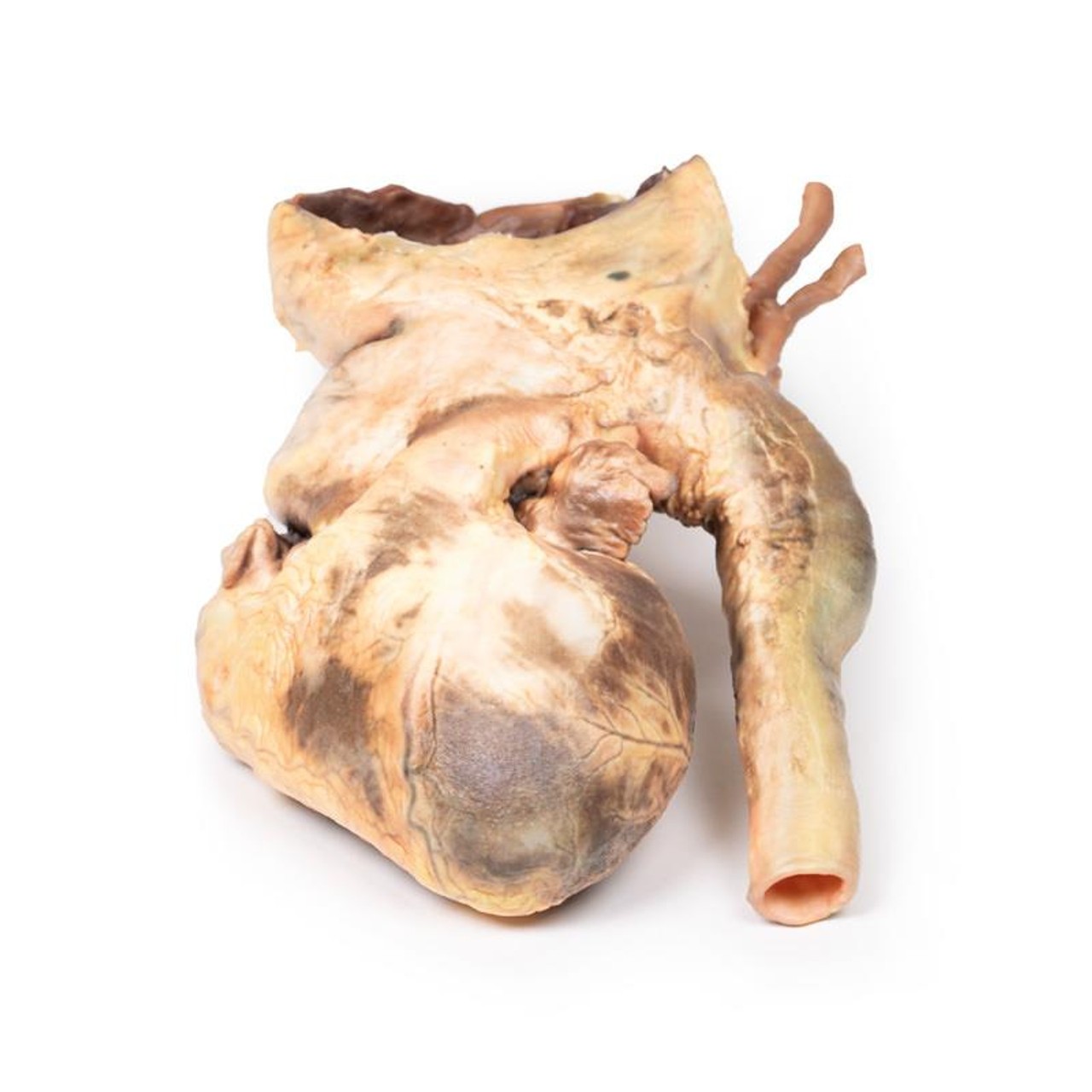
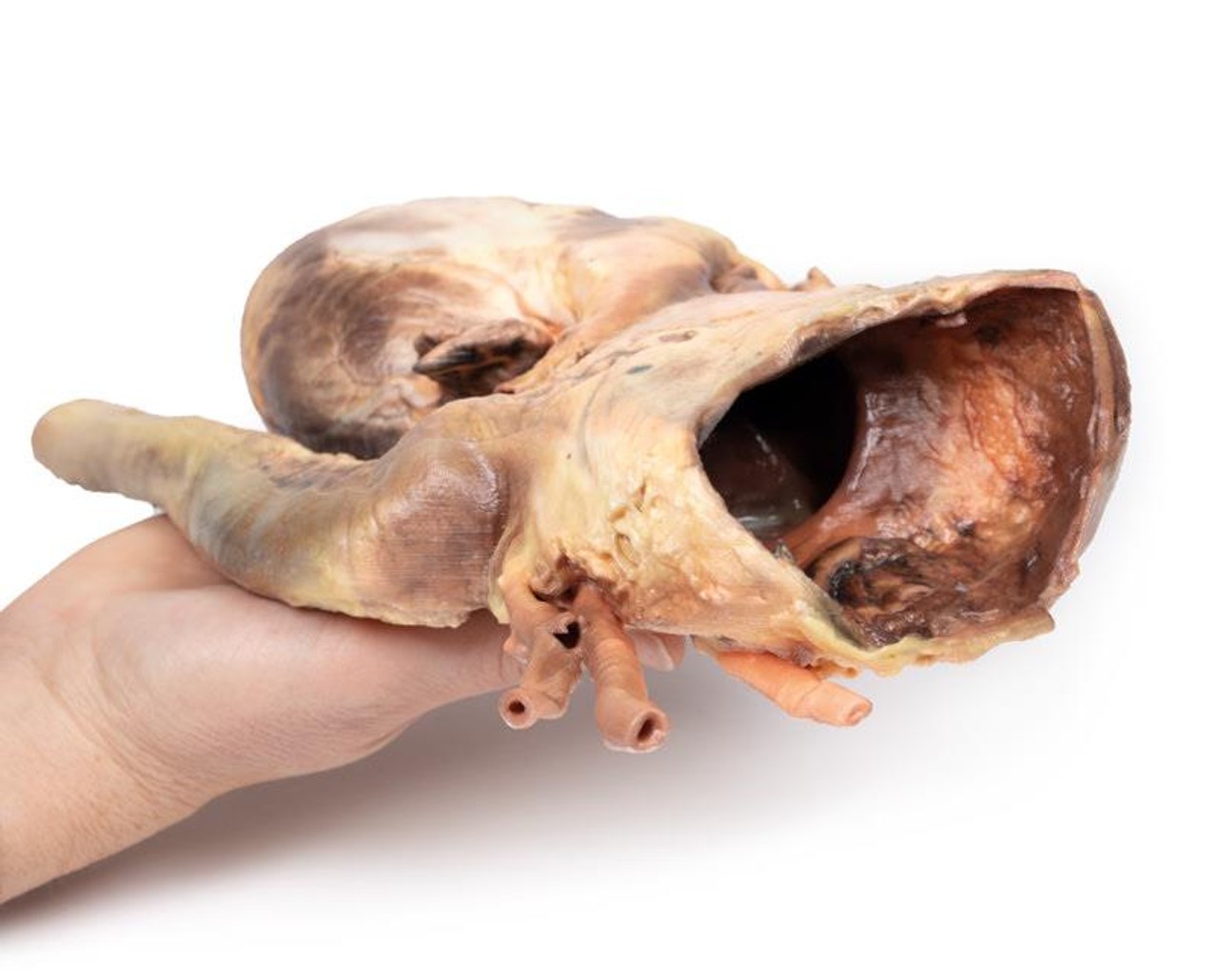
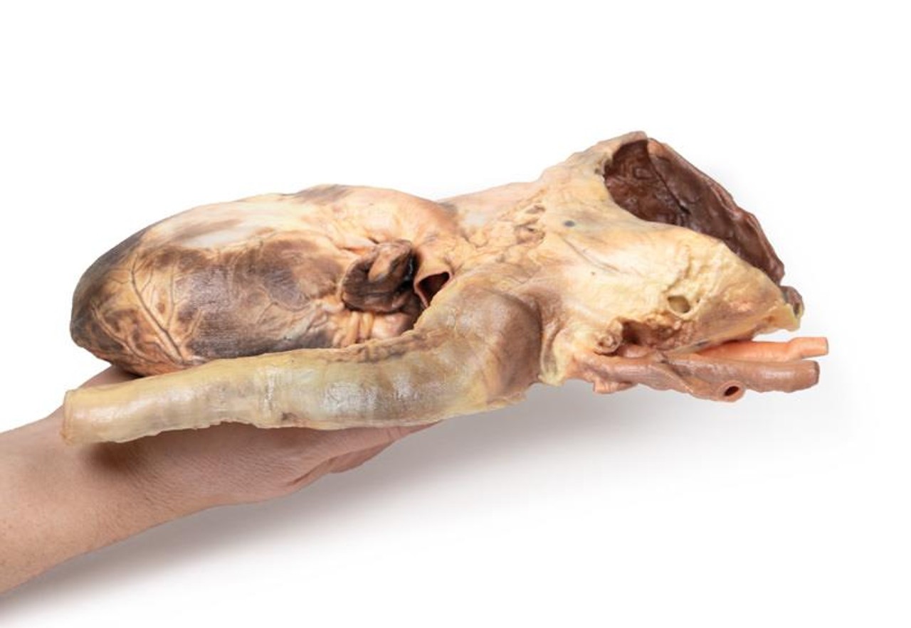
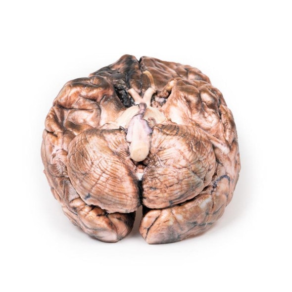
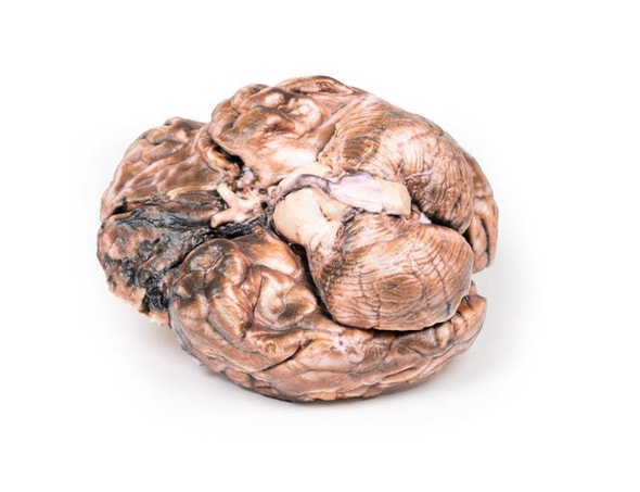
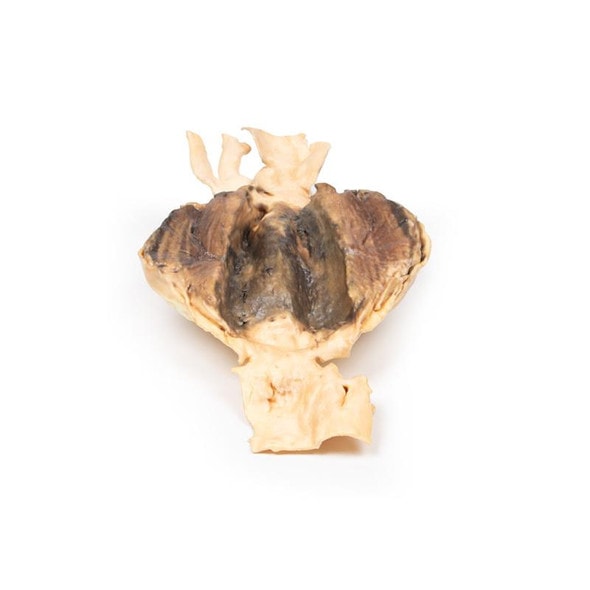
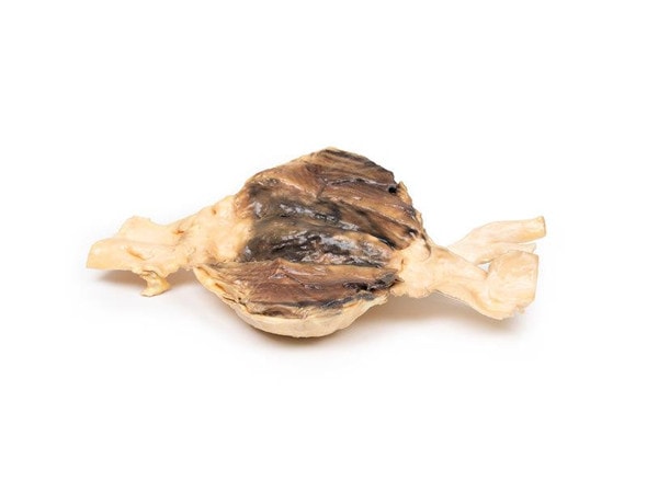
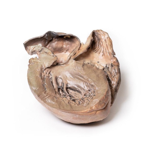
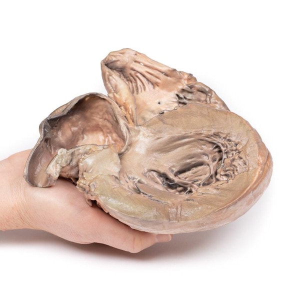
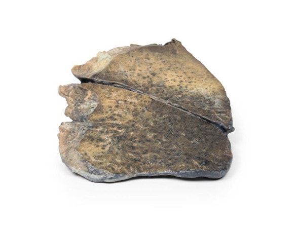
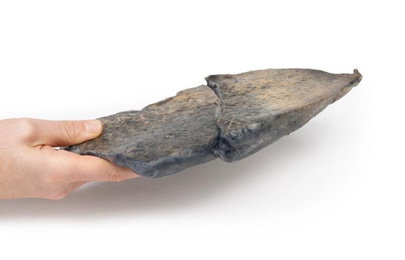
This specimen is the patient's enlarged heart, including the aortic arch and descending aorta. The ascending aorta is dilated up to 7 cm in diameter, and is expanded superiorly by a large aneurysmal bulge 11 x 13 cm in diameter. This has been opened to display the wrinkled scarred intimal surface. There is also marked atheroma of the intima. The innominate, left common carotid and subclavian arteries have been displaced towards the patients left by the aneurysm. On the internal surface of the aneurysm there is a ridge-like thickening 5 mm high. This is the site of attachment of the pericardial sac externally. There is marked congestion of small blood vessels in the adventitia of the aorta. This is a syphilitic aneurysm of the arch of the aorta.
Further Information
Syphilis is a chronic infection caused by the spirochete Treponema pallidum. Sexually transmitted infection is most common but it may also be congenitally acquired by transplacental transmission of the bacteria. Those who have the higher risk of syphilis infection include those of a sexually active age, intravenous drug user, HIV-infected patients and male same sex relationships. Syphilis infection rates decreased significantly with the introduction of penicillin in 1943; it remains the main treatment today. However, the infection rate has been increasing since the early 2000s.
Syphilis is divided into three clinical stages with distinct clinical and pathological features with characteristic proliferative endarteritis affecting small vessels.
Primary syphilis occurs usually 3 weeks after initial infection. This manifests typically as a single, painless and erythematous lesion called a chancre at the site of inoculation. The syphilis spreads throughout the body from this chancre which then heals spontaneously after 3 to 6 weeks.
Secondary syphilis occurs weeks to a few months after the primary chancre resolves in 75% of untreated patients. During this stage patients commonly have generalized symptoms, such as malaise and lymphadenopathy and skin rashes. Palmar/plantar rashes are the most frequent site but rashes can be diffuse. These rashes can be maculopapular, scaly or pustular.
Condylomata lata are elevated gray plaques that arise on the moist mucous membranes such as oral or genital regions. Other less common manifestations include hepatitis, gastrointestinal invasion or ulceration and neurosyphilis - discussed below.
Tertiary syphilis has three main characteristics: cardiovascular syphilis, neurosyphilis and gummatous syphilis. These occur after a latent period of 5 years or more in ? of untreated patients. Cardiovascular syphilis involves an aortitis for which the exact pathophysiology is unclear. The vasculitis involves the ascending thoracic aorta leading to progressive dilation of the aortic root. This can lead to aortic valve insufficiency from dilation of the aortic valve ring. Endarteritis of the vasa vasorum leads to scarring
of the media with loss of muscle and elastic tissue leading to the formation of aneurysms. Clinical manifestation usually happens 15-30 years post initial infection.
Neurosyphilis can be symptomatic or asymptomatic. It occurs in 10% of untreated patients. Early clinical manifestations include headaches, meningitis, hearing loss and ocular involvement, most commonly uveitis, causing vision loss. Late manifestations can occur up to 25 years post initial infection. Main features are meningovascular neurosyphilis, paretic neurosyphilis and tabes dorsalis. Meningovascular involvement involves chronic meningitis and endarteritis which can lead to strokes. Tabes dorsalis is caused from degeneration of the posterior columns within the spinal cord. This causes loss of proprioception, ataxia, loss of pain sensation, and loss of reflexes. Paretic neurosyphilis is caused by invasion and damage of the brain parenchyma, most commonly the frontal lobes. This leads to progressive cognitive impairment and mood disturbance.
Gummatous syphilis is characterized by the formation of nodular lesions most commonly bone, skin and mucosa of the upper airway and mouth called gummas. These can occur anywhere including viscera. The formation of gummas is rare but occurs more frequently in HIV-infected patients. Skeletal involvement causes pain and pathological fractures.
Advantages of 3D Printed Anatomical Models
- 3D printed anatomical models are the most anatomically accurate examples of human anatomy because they are based on real human specimens.
- Avoid the ethical complications and complex handling, storage, and documentation requirements with 3D printed models when compared to human cadaveric specimens.
- 3D printed anatomy models are far less expensive than real human cadaveric specimens.
- Reproducibility and consistency allow for standardization of education and faster availability of models when you need them.
- Customization options are available for specific applications or educational needs. Enlargement, highlighting of specific anatomical structures, cutaway views, and more are just some of the customizations available.
Disadvantages of Human Cadavers
- Access to cadavers can be problematic and ethical complications are hard to avoid. Many countries cannot access cadavers for cultural and religious reasons.
- Human cadavers are costly to procure and require expensive storage facilities and dedicated staff to maintain them. Maintenance of the facility alone is costly.
- The cost to develop a cadaver lab or plastination technique is extremely high. Those funds could purchase hundreds of easy to handle, realistic 3D printed anatomical replicas.
- Wet specimens cannot be used in uncertified labs. Certification is expensive and time-consuming.
- Exposure to preservation fluids and chemicals is known to cause long-term health problems for lab workers and students. 3D printed anatomical replicas are safe to handle without any special equipment.
- Lack of reuse and reproducibility. If a dissection mistake is made, a new specimen has to be used and students have to start all over again.
Disadvantages of Plastinated Specimens
- Like real human cadaveric specimens, plastinated models are extremely expensive.
- Plastinated specimens still require real human samples and pose the same ethical issues as real human cadavers.
- The plastination process is extensive and takes months or longer to complete. 3D printed human anatomical models are available in a fraction of the time.
- Plastinated models, like human cadavers, are one of a kind and can only showcase one presentation of human anatomy.
Advanced 3D Printing Techniques for Superior Results
- Vibrant color offering with 10 million colors
- UV-curable inkjet printing
- High quality 3D printing that can create products that are delicate, extremely precise, and incredibly realistic
- To improve durability of fragile, thin, and delicate arteries, veins or vessels, a clear support material is printed in key areas. This makes the models robust so they can be handled by students easily.

