Description
Developed from real patient case study specimens, the 3D printed anatomy model pathology series introduces an unmatched level of realism in human anatomy models. Each 3D printed anatomy model is a high-fidelity replica of a human cadaveric specimen, focusing on the key morbidity presentations that led to the deceasement of the patient. With advances in 3D printing materials and techniques, these stories can come to life in an ethical, consistently reproduceable, and easy to handle format. Ideal for the most advanced anatomical and pathological study, and backed by authentic case study details, students, instructors, and experts alike will discover a new level of anatomical study with the 3D printed anatomy model pathology series.
Clinical History
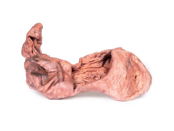
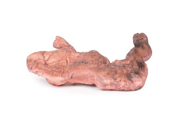
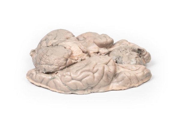
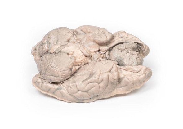
A 62-year-old woman presented with disorientation to time, place, and person. Physical examination revealed no localized neurological signs. Radiological investigations revealed a space occupying lesion in the floor of the 3rd ventricle. The tissue was removed during surgery, but the lesion could not be completely excised. Histology confirmed the diagnosis of Craniopharyngioma. Post-operatively the patient developed complex metabolic disturbances, probably hypothalamic in origin. She gradually deteriorated, and 10 weeks after admission she died following an episode of gastric aspiration.
Pathology
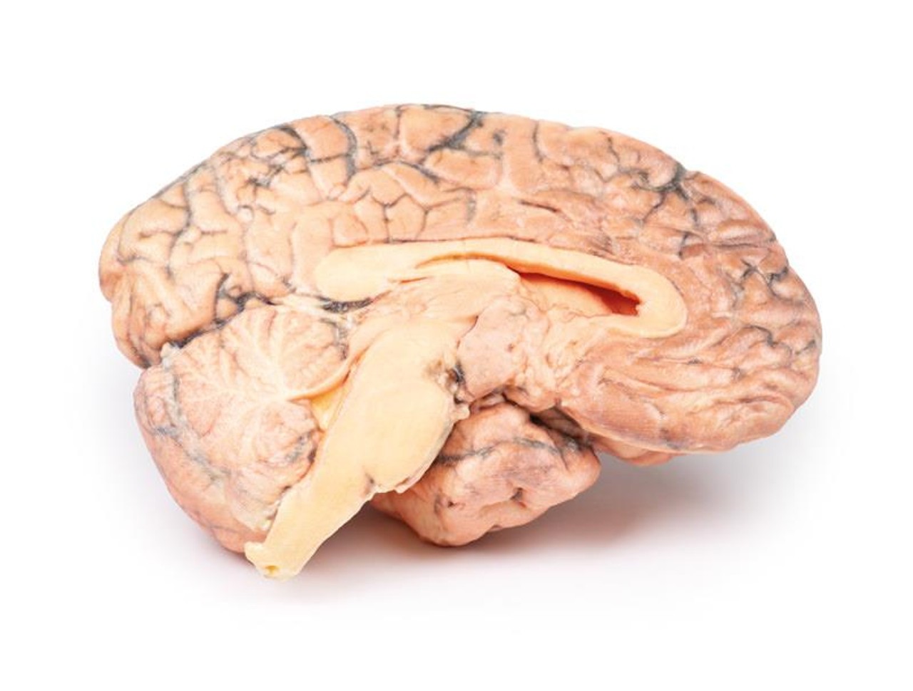
The brain has been sectioned in the sagittal plane, displaying the medial surface. A pink-grey, ovoid tumor measuring 2.5 x 1.5 cm on the cut surface is centered in the region of the hypothalamus. It is encapsulated except at its ventral pole where tissue has been removed at previous surgery, and the cut surface reveals a microcystic or spongy appearance. The tumor distorts the third ventricle and extends to obliterate the Foramen of Munro. The optic chiasm is displaced caudally (arrow). Previous ventriculo-atrial shunting has prevented dilatation of the lateral ventricles despite this obstruction.
Further Information
Craniopharyngiomas constitute 1-3% of all brain tumors, and 5-10% in children, with a bimodal distribution favoring ages 5-14 years, and a second peak between ages 50-75 years. There is a higher incidence in Japan and parts of Africa. Craniopharyngiomas are epithelial tumors generally arising from the pituitary stalk. Other sites of origin include the sella turcica, optic system and third ventricle. There are frequently solid and cystic components, the latter containing cholesterol crystals. Craniopharyngiomas can be divided into two categories - adamantinomatous and papillary types, each with distinct histology and genetic alterations, although the prognostic significance of these types remains unclear.
Treatment includes surgical resection and radiation therapy (RT) to treat and post-surgical residual disease. Prognosis depends largely on tumor resection, control and treatment-related complications arising from local and endocrine and local sequelae.
Advantages of 3D Printed Anatomical Models
- 3D printed anatomical models are the most anatomically accurate examples of human anatomy because they are based on real human specimens.
- Avoid the ethical complications and complex handling, storage, and documentation requirements with 3D printed models when compared to human cadaveric specimens.
- 3D printed anatomy models are far less expensive than real human cadaveric specimens.
- Reproducibility and consistency allow for standardization of education and faster availability of models when you need them.
- Customization options are available for specific applications or educational needs. Enlargement, highlighting of specific anatomical structures, cutaway views, and more are just some of the customizations available.
Disadvantages of Human Cadavers
- Access to cadavers can be problematic and ethical complications are hard to avoid. Many countries cannot access cadavers for cultural and religious reasons.
- Human cadavers are costly to procure and require expensive storage facilities and dedicated staff to maintain them. Maintenance of the facility alone is costly.
- The cost to develop a cadaver lab or plastination technique is extremely high. Those funds could purchase hundreds of easy to handle, realistic 3D printed anatomical replicas.
- Wet specimens cannot be used in uncertified labs. Certification is expensive and time-consuming.
- Exposure to preservation fluids and chemicals is known to cause long-term health problems for lab workers and students. 3D printed anatomical replicas are safe to handle without any special equipment.
- Lack of reuse and reproducibility. If a dissection mistake is made, a new specimen has to be used and students have to start all over again.
Disadvantages of Plastinated Specimens
- Like real human cadaveric specimens, plastinated models are extremely expensive.
- Plastinated specimens still require real human samples and pose the same ethical issues as real human cadavers.
- The plastination process is extensive and takes months or longer to complete. 3D printed human anatomical models are available in a fraction of the time.
- Plastinated models, like human cadavers, are one of a kind and can only showcase one presentation of human anatomy.
Advanced 3D Printing Techniques for Superior Results
- Vibrant color offering with 10 million colors
- UV-curable inkjet printing
- High quality 3D printing that can create products that are delicate, extremely precise, and incredibly realistic
- To improve durability of fragile, thin, and delicate arteries, veins or vessels, a clear support material is printed in key areas. This makes the models robust so they can be handled by students easily.

