Description
Developed from real patient case study specimens, the 3D printed anatomy model pathology series introduces an unmatched level of realism in human anatomy models. Each 3D printed anatomy model is a high-fidelity replica of a human cadaveric specimen, focusing on the key morbidity presentations that led to the deceasement of the patient. With advances in 3D printing materials and techniques, these stories can come to life in an ethical, consistently reproduceable, and easy to handle format. Ideal for the most advanced anatomical and pathological study, and backed by authentic case study details, students, instructors, and experts alike will discover a new level of anatomical study with the 3D printed anatomy model pathology series.
Clinical History
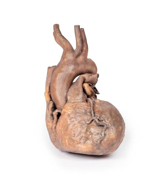
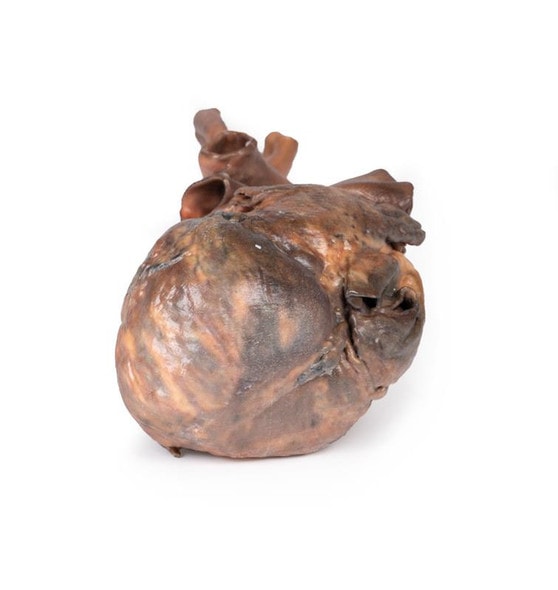
This specimen was found during a routine post-mortem of a 56-year-old male who died of rheumatic heart disease.
Pathology
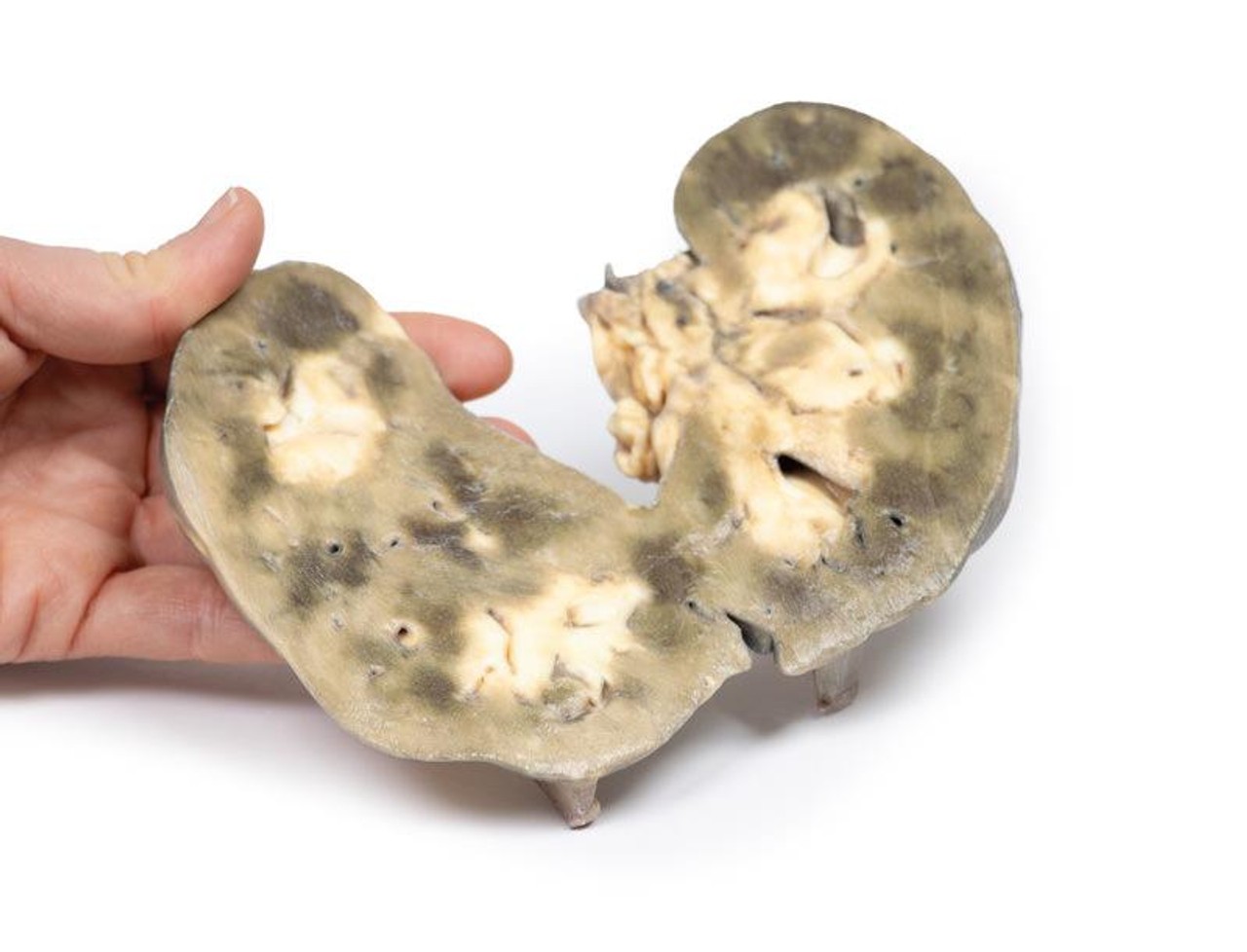
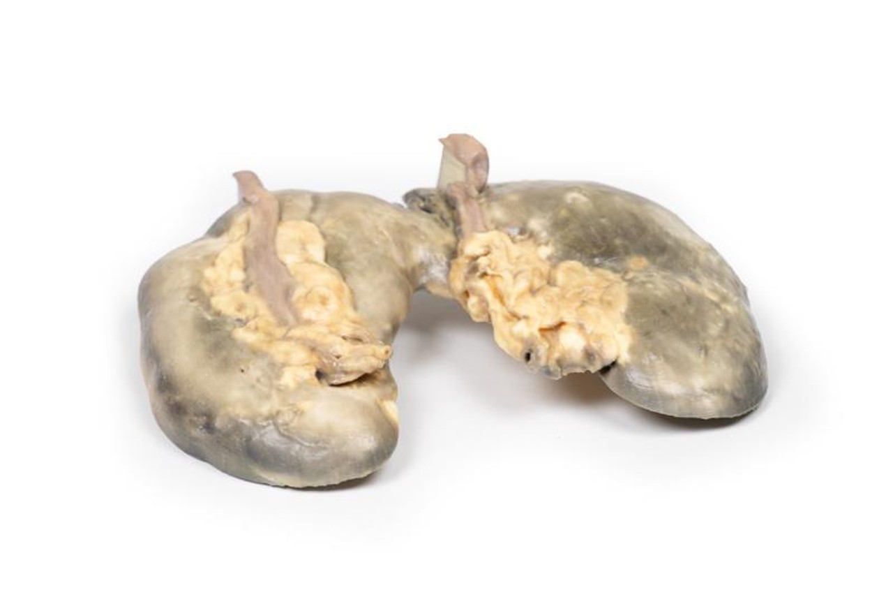
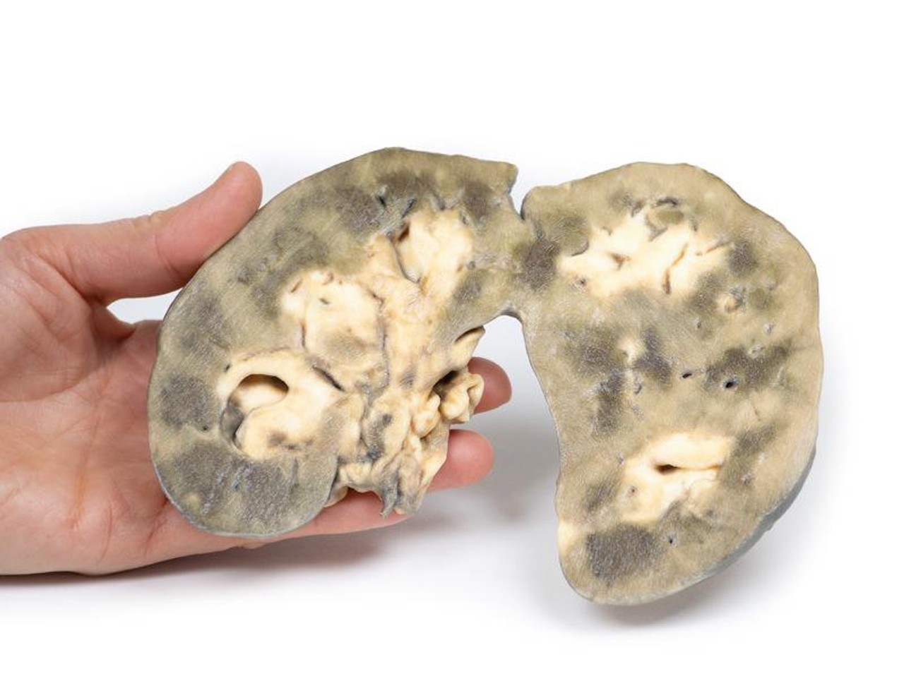
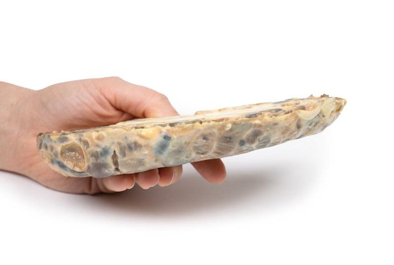
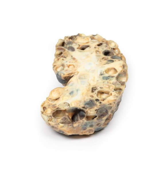
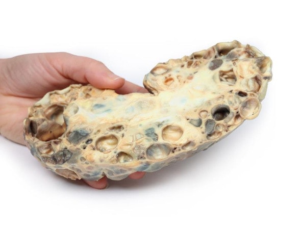
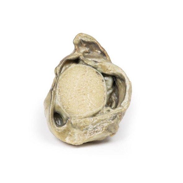
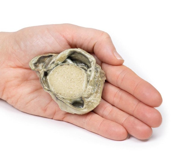
The kidney is 12 cm in length and the two parts are fused at the lower pole forming this horseshoe-like shape. The ureters can be seen emerging from the hilum on the anterior aspect of the 3D print. The kidney is bisected in the horizontal plane which is evident on the posterior aspect. There is persistent fetal lobulation. The renal pelvis is characteristically antero-medial positioned with the ureters travelling anterior to the fused lower poles or isthmus of the kidney.
Further Information
Horseshoe kidneys are the commonest renal developmental abnormality. This anomaly is twice as common in males as in females. They are found in about 1 in 500 to 1000 post-mortem examinations. Most cases are sporadic but may be associated with some chromosomal anomalies, such as those resulting in Downs and Edwards Syndromes as well as non-aneuploidic anomalies, such as VACTERL* association. In 90% of cases an isthmus of renal tissue connects the lower poles of the kidneys across the midline, forming a horseshoe shape. Fusion of the upper lobes is rare. The renal pelvis that drain the two halves of the horseshoe kidney are directed more anteriorly than normal and there is some angulation of ureters as they cross the isthmus.
This malformation is usually asymptomatic and picked up incidentally on routine ultrasound or CT scans. These kidneys usually function normally. There is an increased incidence of urinary calculi, possibly due to angulation of the ureters and to the resulting stasis. There is an increased risk of hydronephrosis usually from pelvi-ureteric junction obstruction. There is a higher incidence of urinary tract infections mainly due to vesico-ureteric reflux. A higher incidence in some forms of renal malignancies (e.g. transitional cell carcinoma and Wilms tumors) has also been described.
(*VACTERL = Vertebral defects, Anal atresia, Cardiac defects, Tracheo-Esophageal fistula, Renal anomalies, and Limb abnormalities).
Advantages of 3D Printed Anatomical Models
- 3D printed anatomical models are the most anatomically accurate examples of human anatomy because they are based on real human specimens.
- Avoid the ethical complications and complex handling, storage, and documentation requirements with 3D printed models when compared to human cadaveric specimens.
- 3D printed anatomy models are far less expensive than real human cadaveric specimens.
- Reproducibility and consistency allow for standardization of education and faster availability of models when you need them.
- Customization options are available for specific applications or educational needs. Enlargement, highlighting of specific anatomical structures, cutaway views, and more are just some of the customizations available.
Disadvantages of Human Cadavers
- Access to cadavers can be problematic and ethical complications are hard to avoid. Many countries cannot access cadavers for cultural and religious reasons.
- Human cadavers are costly to procure and require expensive storage facilities and dedicated staff to maintain them. Maintenance of the facility alone is costly.
- The cost to develop a cadaver lab or plastination technique is extremely high. Those funds could purchase hundreds of easy to handle, realistic 3D printed anatomical replicas.
- Wet specimens cannot be used in uncertified labs. Certification is expensive and time-consuming.
- Exposure to preservation fluids and chemicals is known to cause long-term health problems for lab workers and students. 3D printed anatomical replicas are safe to handle without any special equipment.
- Lack of reuse and reproducibility. If a dissection mistake is made, a new specimen has to be used and students have to start all over again.
Disadvantages of Plastinated Specimens
- Like real human cadaveric specimens, plastinated models are extremely expensive.
- Plastinated specimens still require real human samples and pose the same ethical issues as real human cadavers.
- The plastination process is extensive and takes months or longer to complete. 3D printed human anatomical models are available in a fraction of the time.
- Plastinated models, like human cadavers, are one of a kind and can only showcase one presentation of human anatomy.
Advanced 3D Printing Techniques for Superior Results
- Vibrant color offering with 10 million colors
- UV-curable inkjet printing
- High quality 3D printing that can create products that are delicate, extremely precise, and incredibly realistic
- To improve durability of fragile, thin, and delicate arteries, veins or vessels, a clear support material is printed in key areas. This makes the models robust so they can be handled by students easily.


